
Page 98 - IJB-8-2
P. 98
3D-printed Stent Coated with Dipyridamole-loaded Nanofiber
A B
D
C
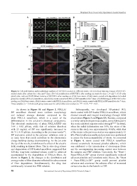
Figure 6. Cell proliferation and morphology analysis of HUVECs seeded on different stents. (A) Live/dead staining images of HUVEC-
seeded stents after culture for 1 day and 7 days. (B) Cell proliferation of HUVECs after seeding on stents for days 1, 4 and 7. (C) F-actin
(phalloidin, red) and DAPI (blue) staining of HUVECs after seeding on (C(i)) bare stents, (C(ii)) stents coated with dipyridamole-loaded
poly(D,L-lactide) (PDLLA) nanofibers, and (C(iii)) stents coated with PDLLA/DP nanofibers for 7 days. (D) SEM images of HUVECs after
seeding on (D(i)) bare stents, (D(ii)) stents coated with PDLLA nanofibers, and (D(iii)) stents coated with PDLLA/DP nanofibers for 7 days.
Three samples (n = 3) from each group were used for cell proliferation evaluation. *P < 0.05, **P < 0.01.
As shown in Figure S1 and Figure 2, PDLLA/ Subsequently, we developed 3D-printed PCL
DP nanofibers showed more uniform morphology stents coated with DP-loaded PDLLA nanofibers, which
and reduced average diameter compared to the showed smooth and integral morphology through SEM
plain PDLLA nanofibers, which is a result of the observation (Figure 3 and Figure S3). Besides, compared
differentiation in the solution’s electrical conductivity. to similar stents in the previous studies, stents fabricated in
The electrical conductivity of plain PDLLA/HFIP was this work exhibited superior radial strength [38,39] . In detail,
3.64 ± 0.02 μS/cm, while that of solution dissolved when the radial deformation was 50%, the stress of the
with 25 mg/mL of DP was significantly increased to stents in this study was approximately 40 kPa, while that
68.71 ± 0.39 μS/cm. According to the previous works , of the stents in the previous studies was approximately 15
[37]
DP molecules added in the polymer solutions exist in kPa. Platelet adhesion and hemolysis tests were performed
an ionic form that could contribute to the electrostatic to assess the hemocompatibility of stents. As presented
charge build-up during electrospinning. As the jet exits in Figure 4, stents coated with PDLLA nanofibers
the tip of the nozzle, it reinforces the effect of the electric showed extensively increased platelet adhesion, which
field, resulting in thinner fibers. The in vitro drug release was attributed to the introduction of electrospun fibers
and degradation of DP-loaded nanofibers suggested that and the accompanying increasing contact area between
120-day long-term sustained release could be achieved the samples and PRP solution. Conversely, the “Stent +
accompanied by the degradation of PDLLA fibers. As Nano-PDLLA-DP” group showed significantly reduced
shown in Figure 2, the changes in the distribution and adherent platelets and hemolysis ratio. Hence, the “Stent
average value of fiber diameter reflected the advancement + Nano-PDLLA-DP” group could prevent platelet
of fiber degradation. Simultaneously, the drug was adhesion and counteract the adverse effect of increased
continuously released into PBS solution. contact area due to the benefits of DP.
90 International Journal of Bioprinting (2022)–Volume 8, Issue 2