
Page 100 - IJB-8-2
P. 100
3D-printed Stent Coated with Dipyridamole-loaded Nanofiber
coated with PDLLA/DP nanofibers could inhibit the
proliferation of SMC and had no detrimental effects
on endothelial cells in vitro. Furthermore, the in vivo
A B
implantation of stents coated with PDLLA/DP nanofibers
showed initial patency and continuous endothelialization
and alleviated neointimal formation compared to bare
stents. Collectively, the integrated stents coated with DP-
loaded PDLLA nanofibers showed great potential for
restenosis prevention and endothelialization. In future
research, the decrease in strut thickness and the long-term
in vivo implantation of improved stents will be further
explored.
Acknowledgments
The authors acknowledge funding support from the
Tsinghua University Initiative Scientific Research
Program (20197050024) and the 111 Project (B17026).
Conflicts of interest
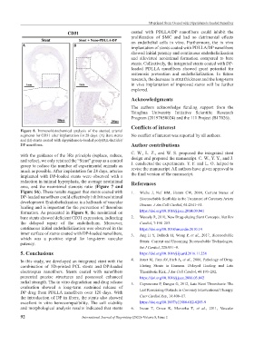
Figure 8. Immunohistochemical analysis of the stented arterial
segments for CD31 after implantation for 28 days. (A) Bare stents No conflict of interest was reported by all authors.
and (B) stents coated with dipyridamole-loaded poly(D,L-lactide)/
DP nanofibers. Author contributions
with the guidance of the 3Rs principle (replace, reduce, C. W., L. Z., and W. S. proposed the integrated stent
and refine), we only retained the “Stent” group as a control design and prepared the manuscript. C. W., Y. Y., and J.
group to reduce the number of experimental animals as J. conducted the experiments. Y. F. and L. O. helped to
revise the manuscript. All authors have given approval to
much as possible. After implantation for 28 days, arteries
implanted with DP-loaded stents were observed with a the final version of the manuscript.
reduction in intimal hyperplasia, the average neointimal References
area, and the neointimal stenosis ratio (Figure 7 and
Figure S6). These results suggest that stents coated with 1. Wiebe J, Nef HM, Hamm CW, 2014, Current Status of
DP-loaded nanofibers could effectively inhibit neointimal Bioresorbable Scaffolds in the Treatment of Coronary Artery
development. Endothelialization is a hallmark of vascular Disease. J Am Coll Cardiol, 64:2541–51.
healing and is important for the prevention of thrombus
formation. As presented in Figure 8, the neointimal on https://doi.org/10.1016/j.jacc.2014.09.041
bare stents showed deficient CD31 expression, indicating 2. Wessely R, 2010, New Drug-eluting Stent Concepts. Nat Rev
the delayed repair of the endothelium. Moreover, Cardiol, 7:194–203.
continuous initial endothelialization was observed in the https://doi.org/10.1038/nrcardio.2010.14
inner surface of stents coated with DP-loaded nanofibers, 3. Ang H Y, Bulluck H, Wong P, et al., 2017, Bioresorbable
which was a positive signal for long-term vascular Stents: Current and Upcoming Bioresorbable Technologies.
patency.
Int J Cardiol, 228:931–9.
5. Conclusions https://doi.org/10.1016/j.ijcard.2016.11.258
In this study, we developed an integrated stent with the 4. Joner M, Finn AV, Farb A, et al., 2006, Pathology of Drug-
combination of 3D-printed PCL stents and DP-loaded Eluting Stents in Humans. Delayed Healing and Late
electrospun nanofibers. Stents coated with nanofibers Thrombotic Risk. J Am Coll Cardiol, 48:193–202.
presented precise structures and possessed enhanced https://doi.org/10.1016/j.jacc.2006.03.042
radial strength. The in vitro degradation and drug release 5. Capranzano P, Dangas G, 2012, Late Stent Thrombosis: The
evaluation showed a long-term sustained release of Last Remaining Obstacle in Coronary Interventional Therapy.
DP drug from PDLLA nanofibers over 120 days. With
the introduction of DP in fibers, the stents also showed Curr Cardiol Rep, 14:408–17.
excellent in vitro hemocompatibility. The cell viability https://doi.org/10.1007/s11886-012-0283-9
and morphological analysis results indicated that stents 6. Inoue T, Croce K, Morooka T, et al., 2011, Vascular
92 International Journal of Bioprinting (2022)–Volume 8, Issue 2