
Page 99 - IJB-8-2
P. 99
Wang, et al.
A B C D
E F G
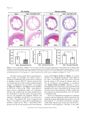
Figure 7. In vivo implantation evaluation of the bare stents and stents coated with dipyridamole-loaded poly(D,L-lactide) (PDLLA)/DP
nanofibers. H&E staining images of (A) bare stents and (B) stents coated with PDLLA/DP nanofibers. Masson staining images of (C) bare
stents and (D) stents coated with PDLLA/DP nanofibers. Quantitative analysis of (C) neointimal thickness, (D) neointimal area, and (E)
neointimal stenosis ratio. Three samples (n = 3) from each group were used to analyze neointimal development *P < 0.05.
The native arteries consist of three structural layers: release of DP (Figure 5C(iii) and 5D(iii)). In contrast,
intima, media, and adventitia. The intima includes a HUVECs were adequately overspread on the surface of
monolayer of endothelial cells, and the media is composed the “Stent + Nano-PDLLA-DP” group, which indicated
of abundant vascular SMCs . Adverse reactions after that the release of DP showed no adverse effects on
[40]
stent implantation are closely related to cellular biological the cell proliferation and morphology of endothelial
changes in endothelial cells and SMCs. As presented cells (Figure 6C(iii) and 6D(iii)). Therefore, it can be
in Figure S5, Figure 5, and Figure 6, both RASMCs determined that 3D-printed stents coated with DP-loaded
and HUVECs seeded on the “Stent + Nano-PDLLA” nanofibers (the “Stent + Nano-PDLLA-DP” group) could
group showed a significantly higher cell proliferation obviously prevent the proliferation of SMCs seeded onto
rate than cells seeded on the bare stents, benefiting from stents and have no detrimental effects on the cell viability
the introduction of nanofibers and an increased contact of endothelial cells.
area for cell attachment and migration. Different cell The in vivo implantation assessment was conducted
behaviors of RASMCs and HUVECs were observed in to further verify the performance of DP-loaded stents. The
the “Stent + Nano-PDLLA-DP” group. Compared to the detriments of stents without drug delivery have been well
“Stent + Nano-PDLLA” group, the cell proliferation of demonstrated [1-3,8,9] . We believe that PDLLA nanofiber-
RASMCs seeded on the “Stent + Nano-PDLLA-DP” coated stents without drugs cannot cope with the above-
group showed an obvious decrease due to the continuous mentioned intimal hyperplasia. Therefore, in this study,
International Journal of Bioprinting (2022)–Volume 8, Issue 2 91