
Page 192 - IJB-8-3
P. 192
Bioprinting of a Hepatic Tissue Model
A
B
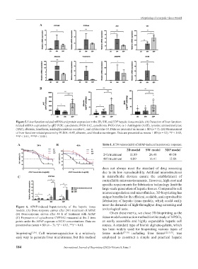
Figure 5. Liver function-related mRNA and protein expression in the 2D, SW, and 3DP hepatic tissue models. (A) Detection of liver function-
related mRNA expression by qRT-PCR: cytochrome P450-1A2, cytochrome P450-3A4, α-1-Antitrypsin (AAT), tyrosine aminotransferase
(TAT), albumin, transferrin, asialoglycoprotein receptor-1, and cytokeratin-18. Data are presented as means ± SD (n = 3). (B) Measurement
of liver function-related proteins by ELISA: AAT, albumin, and blood urea nitrogen. Data are presented as means ± SD (n = 12). *P < 0.05,
**P < 0.01, ***P < 0.001.
A B Table 1. IC50 values (mM) of APAP-induced hepatotoxic response.
2D model SW model 3DP model
24 h treatment 11.89 26.48 49.54
48 h treatment 0.89 16.41 12.88
does not always meet the standard of drug screening
due to its low reproducibility. Artificial microstructures
C in microfluidic devices ensure the establishment of
controllable microenvironments. However, high cost and
specific requirements for fabrication technology limit the
large-scale generation of hepatic tissues. Compared to cell
microencapsulation and microfluidics, 3D bioprinting has
unique benefits for the efficient, scalable, and reproducible
fabrication of hepatic tissue models, which could easily
meet the demands of high-throughput drug screening and
Figure 6. APAP-induced hepatotoxicity of the hepatic tissue
models. (A) Dose-response curves after 24 h treatment of APAP. toxicological tests.
(B) Dose-response curves after 48 h of treatment with APAP. Given these merits, we chose 3D bioprinting as the
(C) Expression of cytochrome CYP1A2 measured at the 2 time tissue model construction method for this study of hiPSCs,
points under the APAP exposure of IC50 concentrations. Data are an easily accessible and highly expandable hepatic cell
presented as means ± SD (n = 3). *P < 0.05, **P < 0.01. source. A standard type of bioink alginate-gelatin, which
has been widely used for bioprinting various types of
bioprinting [3,36] . Cell microencapsulation is a relatively tissue models [37-39] including liver tissue [12,13,18] , was
easy way to generate liver microtissues, but this method employed to construct a simple and practical hepatic
184 International Journal of Bioprinting (2022)–Volume 8, Issue 3