
Page 191 - IJB-8-3
P. 191
He, et al.
A
B
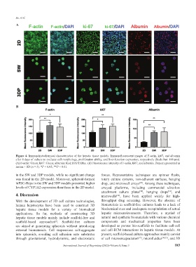
Figure 4. Immunohistochemical characteristics of the hepatic tissue models. Immunofluorescent images of F-actin, ki67, and albumin
after 8 days of culture to evaluate cell morphology, proliferation ability, and liver-function expression, respectively (Scale bar: 100 μm).
(A) F-actin: Green; ki67: Green; albumin: Red; DAPI: Blue. (B) Fluorescence intensity of F-actin, ki67, and albumin. Data are presented as
means ± SD (n = 3). *P < 0.05, **P < 0.01.
in the SW and 3DP models, while no significant change forces. Representative techniques are spinner flasks,
was found in the 2D model. Moreover, spheroid-formed rotary culture systems, non-adherent surfaces, hanging
hiPSC-Heps in the SW and 3DP models presented higher drop, and microwell arrays . Among these techniques,
[29]
levels of CYP1A2 expression than those in the 2D model. arrayed platforms, including commercial ultra-low
attachment culture plates , hanging drops , and
[31]
[30]
4. Discussion microwells , have been applied widely for high-
[32]
With the development of 3D cell culture technologies, throughput drug screening. However, the absence of
human hepatocytes have been used to construct 3D biomaterials in scaffold-free cultures leads to a lack of
hepatic tissue models for a variety of biomedical biochemical cues and inadequate recapitulation of actual
applications. So far, methods of constructing 3D hepatic microenvironments. Therefore, a myriad of
hepatic tissue models mainly include scaffold-free and natural and synthetic biomaterials with various chemical
scaffold-based approaches . Scaffold-free cultures components and mechanical properties have been
[6]
are aimed at generating spheroids without introducing developed as porous bio-scaffolds to facilitate cell-cell
external biomaterials. Cell suspensions self-aggregate and cell-ECM interactions in hepatic tissue models. At
into spheroids, avoiding cell adhesion onto substrates present, scaffold-based culture approaches mainly consist
through gravitational, hydrodynamic, and electrostatic of cell microencapsulation , microfluidics [34,35] , and 3D
[33]
International Journal of Bioprinting (2022)–Volume 8, Issue 3 183