
Page 253 - IJB-8-3
P. 253
Li, et al.
A B C
D E F
G H I
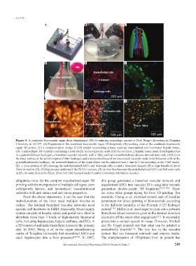
Figure 9. A combined four-nozzle organ three-dimensional (3D) bioprinting technology created at Prof. Wang’s laboratory in Tsinghua
University in 2013 : (A) Equipment of the combined four-nozzle organ 3D bioprinter, (B) working state of the combined four-nozzle
[66]
organ 3D printer, (C) a computer-aided design (CAD) model representing a large scale-up vascularized and innervated hepatic tissue,
(D) a semi-ellipse 3D construct containing a poly (lactic acid-co-glycolic acid) (PLGA) overcoat, a hepatic tissue made from hepatocytes
in a gelatin/chitosan hydrogel, a branched vascular network with a fully confluent endothelialized adipose-derived stem cells (ASCs) on
the inner surface of the gelatin/alginate/fibrin hydrogel, and a hierarchical neural (or innervated) network made from Schwann cells in the
gelatin/hyaluronate hydrogel, the maximal diameter of the semi-ellipse can be adjusted from 1 mm to 2 cm according to the CAD model,
(E) a cross-section of (D) showing the endothelialized ASCs and Schwann cells around a branched channel, (F) a large bundle of nerve
fibers formed in (D), (G) hepatocytes underneath the PLGA overcoat, (H) an interface between the endothelialized ASCs and Schwann cells
in (D), (I) some thin nerve fibers. (from ref. [66] licensed under Creative Commons Attribution license).
ubiquitous roles for the complex vascularized organ 3D this group generated a branched vascular network and
printing with the incorporation of multiple cell types, stem transformed ASCs into vascular ECs using their second-
cells/growth factors, and hierarchical vascular/neural generation double-nozzle 3D bioprinter [50,99-104] . There
networks with anti-suture and anti-stress properties. are some other groups trying for liver 3D printing. For
From the above statements, it can be seen that the example, Chang et al. provided several sets of baseline
multi-functions of the liver need multiple nozzles to parameters for direct printing of biomaterials according
realize. The internal branched vascular networks need to the different humidity of the Pluronic F127 hydrogel
specific soft hardware to fulfill. Especially, blood supply system [114] . Miller et al. used sugar to print out a network
system consists of hepatic artery and portal vein. How to that allows blood vessels to grow in the desired direction
distribute more than 3 kinds of high-density functional and melts off the stents after angioplasty [115] . It eventually
cells, including hepatocytes, hepatic stellates, and ECs, in grows into a conduit system in tissue. However, Birchall
a 3D construct and make them form tissues is the primary and De Coppi pointed out that what can be printed is
task. In 2005, Wang et al. in the organ manufacturing undoubtedly feasible [116] . The key lies in the vascular
center of Tsinghua University first assembled ASCs and system that can transport nutrients and remove waste.
adult hepatocytes into a liver precursor [86-98] . In 2007, The transplantation of 3D-printed liver in patient has
International Journal of Bioprinting (2022)–Volume 8, Issue 3 245