
Page 45 - IJB-8-3
P. 45
Duan, et al.
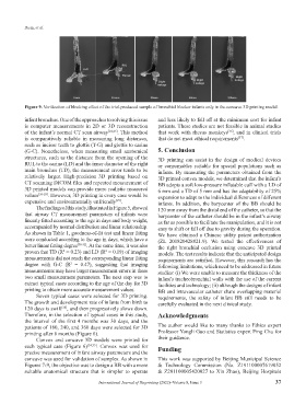
Figure 9. Verification of blocking effect of the trial-produced sample of bronchial blocker infants only in the concave 3D printing model.
infant bronchus. One of the approaches to solving this issue and less likely to fall off at the minimum cost for infant
is computer measurements in 2D or 3D reconstruction patients. These studies are not feasible in animal studies
of the infant’s normal CT scan airway [24,25] . This method that work with rhesus monkeys , and in clinical trials
[36]
is comparatively reliable in measuring long distances, that do not meet ethical requirements .
[37]
such as incisor teeth to glottis (T-G) and glottis to carina
(G-C). Nonetheless, when measuring small anatomical 5. Conclusion
structures, such as the distance from the opening of the 3D printing can assist in the design of medical devices
RUL to the carina (LD) and the inner diameter of the right or consumables suitable for special populations such as
main bronchus (TD), the measurement error tends to be infants. By measuring the parameters obtained from the
relatively larger. High-precision 3D printing based on 3D printed convex models, we determined that the infant’s
CT scanning DICOM files and repeated measurement of BB adopts a soft low-pressure inflatable cuff with a LD of
3D printed models can provide more realistic measured 6 mm and a TD of 5 mm and has the adaptability of 25%
values [26-28] . However, 3D printing in every case would be expansion to adapt to the individual differences of different
expensive and environmentally unfriendly . infants. In addition, the barycenter of the BB should be
[29]
The findings of this study, illustrated in Figure 5, showed 120 mm away from the distal end of the catheter, so that the
that airway CT measurement parameters of infants were barycenter of the catheter should be in the infant’s airway
linearly fitted according to the age in days and body weight, as far as possible to facilitate the manipulation, and it is not
accompanied by normal distribution and linear relationship. easy to shift or fall off due to gravity during the operation.
As shown in Table 1, goodness-of-fit test and linear fitting We have obtained a Chinese utility patent authorization
were conducted according to the age in days, which have a (ZL 201820428821.9). We tested the effectiveness of
better linear fitting degree [30-32] . At the same time, it was also the right bronchial occlusion using concave 3D printed
proven that TD (R = 0.23) and LD (R = 0.19) of imaging models. The test results indicate that the anticipated design
2
2
measurements did not reach the corresponding linear fitting requirements are satisfied. However, this research has the
degree with G-C (R = 0.47), suggesting that imaging following limitations, which need to be addressed in future
2
measurements may have larger measurement errors in these studies: (i) We were unable to measure the thickness of the
two small measurement parameters. The next step was to infant’s tracheobronchial walls with the use of the current
extract typical cases according to the age of the day for 3D facilities and technology; (ii) although the designs of infant
printing to obtain more accurate measurement values. BB and intravascular catheter share overlapping material
Seven typical cases were selected for 3D printing. requirements, the safety of infant BB still needs to be
The growth and development rate of infants from birth to carefully evaluated in the next clinical study.
120 days is swift , and then progressively slows down.
[33]
Therefore, in the selection of typical cases in this study, Acknowledgments
the interval of the first 4 months was 30 days, and the
patients of 180, 240, and 360 days were selected for 3D The author would like to many thanks to Ethics expert
printing after 6 months (Figure 6). Professor Yongli Guo and Statistics expert Ping Chu for
Convex and concave 3D models were printed for their guidance.
each typical case (Figure 6) [34,35] . Convex was used for Funding
precise measurement of infant airway parameters and the
concave was used for validation of samples. As shown in This work was supported by Beijing Municipal Science
Figures 7-9, the objective was to design a BB with a more & Technology Commission (No. Z191100007619052
suitable anatomical structure that is simpler to operate & Z201100005420027 to Xin Zhao), Beijing Hospitals
International Journal of Bioprinting (2022)–Volume 8, Issue 3 37