
Page 127 - IJB-8-4
P. 127
Park, et al.
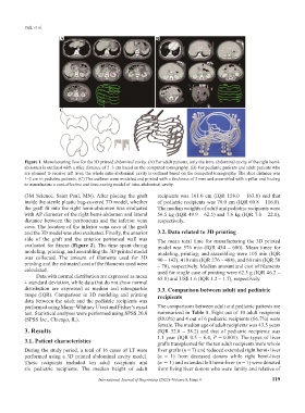
A B
C
Figure 1. Manufacturing flow for the 3D printed abdominal cavity. (A) For adult patients, only the intra-abdominal cavity of the right hemi-
abdomen is outlined with a slice distance of 2–3 cm based on the computed tomography. (B) For pediatric patients and adult patients who
are planned to receive left liver, the whole intra-abdominal cavity is outlined based on the computed tomography. The slice distance was
1–2 cm in pediatric patients. (C) The outlines were modeled and printed with a thickness of 2 mm and assembled with a pillar and footing
to manufacture a cost-effective and time-saving model of intra-abdominal cavity.
(3M Science, Saint Paul, MN). After placing the graft recipients was 161.0 cm (IQR 158.0 – 163.8) and that
inside the sterile plastic bag-covered 3D model, whether of pediatric recipients was 70.0 cm (IQR 60.8 – 116.8).
the graft fit into the right hemi-abdomen was evaluated The median weights of adult and pediatric recipients were
with AP diameter of the right hemi-abdomen and lateral 56.5 kg (IQR 49.9 – 62.5) and 7.6 kg (IQR 7.0 – 22.0),
distance between the peritoneum and the inferior vena respectively.
cava. The location of the inferior vena cava of the graft
and the 3D model was also evaluated. Finally, the anterior 3.2. Data related to 3D printing
side of the graft and the anterior peritoneal wall was The mean total time for manufacturing the 3D printed
evaluated for fitness (Figure 2). The time spent during model was 576 min (IQR 434 – 680). Mean times for
modeling, printing, and assembling the 3D printed model modeling, printing, and assembling were 105 min (IQR
was collected. The amount of filaments used for 3D 90 – 142), 418 min (IQR 276 – 488), and 60 min (IQR 50
printing and the estimated cost of the filaments used were – 70), respectively. Median amount and cost of filaments
calculated. used for single case of printing were 62.5 g (IQR 46.2 –
Data with normal distribution are expressed as mean 65.8) and US$ 1.6 (IQR 1.2 – 1.7), respectively.
± standard deviation, while data that do not show normal
distribution are expressed as median and interquartile 3.3. Comparison between adult and pediatric
range (IQR). Comparison in 3D modeling and printing recipients
data between the adult and the pediatric recipients was
performed using Mann–Whitney U test and Fisher’s exact The comparisons between adult and pediatric patients are
test. Statistical analyses were performed using SPSS 20.0 summarized in Table 1. Eight out of 10 adult recipients
(SPSS Inc., Chicago, IL). (80.0%) and 4 out of 6 pediatric recipients (66.7%) were
female. The median age of adult recipients was 43.5 years
3. Results (IQR 32.8 – 58.2) and that of pediatric recipients was
1.1 year (IQR 0.5 – 6.4, P = 0.001). The types of liver
3.1. Patient characteristics
grafts transplanted for the ten adult recipients were whole
During the study period, a total of 16 cases of LT were liver grafts (n = 7) and reduced extended right hemi- liver
performed using a 3D printed abdominal cavity model. (n = 1) from deceased donors while right hemi-liver
These recipients included ten adult recipients and (n = 1) and extended left hemi-liver (n = 1) were donated
six pediatric recipients. The median height of adult from living liver donors who were family and relative of
International Journal of Bioprinting (2022)–Volume 8, Issue 4 119