
Page 126 - IJB-8-4
P. 126
3D-printed Abdominal Cavity Model for Liver Transplantation
between the recipient’s abdominal cavity and the donor would be placed was masked using Mimics Medical 21.0
liver graft. The most important features of techniques and (Materialise, Leuven, Belgium). For efficient modeling
biomaterials to have for 3D printing of intra-abdominal and printing, the inner surface of the abdominal cavity
cavity are: (i) Harmless to human body since it is used was outlined with a 1 – 3 cm distance between slices.
during surgery, (ii) strong and rigid enough to maintain its While the distance between the printed lines was longer
shape while fitting the liver graft to the 3D printed model, in adult recipients, the distance was relatively shorter in
and (iii) can be readily utilized in emergency operation. pediatric recipients. The anterior wall of the abdominal
Therefore, we decided to use fused deposition modeling cavity was marked on the peritoneal side of the anterior
(FDM)-based 3D printing technique and polylactic acid abdominal wall. The line was continued to the lateral wall
material, and created 3D printed models of LT recipients’ outlining the peritoneal surface. The posterior wall of the
abdominal cavity and utilized them during LT with a abdominal cavity consisted of perirenal fat surrounding
potential for large-for size syndrome. Our 3D model the kidney. The midline of the abdominal cavity was
has advantages of low cost and fast production time outlined along the inferior vena cava and abdominal
compared to previous 3-D printed models used in LT. This aorta. The medial two-third of the anterior wall outline
study is designed to describe methods for manufacturing was removed with a vertical marking that pointed out
3D printed abdominal cavity model and evaluate the the anterior limit of the abdominal cavity. While only the
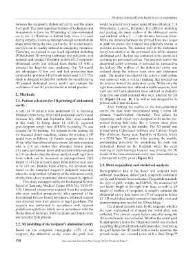
usefulness of our 3D printed model in actual practice. right hemi-abdomen was outlined in adult recipients, both
right and left hemi-abdomen were outlined in pediatric
2. Methods recipients and adult recipient who were planned for left
2.1. Patient selection for 3D printing of abdominal LT (Figure 1A and B). The outline was designed to be
cavity printed with 2 mm thickness.
After marking the outline of the intra-abdominal
A total of 16 patients who underwent LT at Samsung cavity, the data were manipulated using Cinema 4D
Medical Center using 3D printed abdominal cavity model (Maxon, Friedrichsdorf, Germany). Two pillars for
between July 2020 and September 2021 were enrolled supporting each object were designed to fit on the pre-
in this study. In living donor LT (LDLT), expected printed footing that looks like a Korean chess piece
graft-recipient weight ratio (GRWR) exceeding 2% was with a square hole in the middle. The 3D model was
selected for 3D printing. For patients on the waiting list printed using Cubicreator software and Cubicon Single
for deceased donor matching, criteria for printing a 3D Plus (Cubicon, Seong nam, Republic of Korea), which
model were as follows: (i) Female recipient who is not is a FDM Type 3D printer. After printing the models,
10 cm taller than allocated male donor, (ii) male recipient post-printing procedure for assembling the parts was
who is ≥10 cm shorter than allocated female donor, performed. Based on the blueprint where the exact
(iii) same sex between donor and recipient while recipient marking of each footing’s location was printed, the 3D
is ≥10 cm shorter than the donor, and (iv) small right liver model of the intra-abdominal cavity was reassembled on
fossa which can be measured as anteroposterior (AP) a transparent acrylic panel (Figure 1C).
length of ≤13 cm or lateral space from inferior vena cava
to be ≤10 cm. Besides these criteria, the selection was 2.3. Data acquisition and statistical analysis
based on the transplant surgeon’s judgment especially Demographical data of the donor and recipient were
when the recipient had deformity of the abdominal cavity collected. In addition, data of graft, recipients’ abdominal
of which the above-mentioned criteria cannot be applied. cavity, and 3D model were collected. The graft data include
This study was approved by the Institutional Review the type of graft, weight, and GRWR. We measured AP
Board of Samsung Medical Center (IRB No. 2020-07- and lateral length of the right liver fossa as well as AP
118). Informed consent was acquired from the recipients length of midline of recipients to roughly estimate the
who were enrolled prospectively after approval of the abdominal cavity data based on CT of recipients before
Institutional Review Board; for minors, informed consent LT. 3D model data include amount of materials, cost, and
was obtained from their parents or legal guardians. The manufacturing time needed for 3D modeling.
research was performed in accordance with relevant The clinical decision made by the surgeons, whether
guidelines/regulations which are in accordance with the the team maintained or changed the initial plan, was
Declaration of Helsinki. Both recipients and donors were collected. The clinical course before and after using the
not recruited from prisons. 3D printed model was collected. Whether the actual graft
2.2. 3D modeling of the recipient’s abdominal cavity fit appropriately inside the 3D printed model was checked
by putting the graft after back-table procedure. For putting
Based on the computed tomography (CT) of the the graft inside the 3D model with a sterile measure, the
recipient, the abdominal cavity where the graft liver printed model was covered twice with a Steri-Drape™
118 International Journal of Bioprinting (2022)–Volume 8, Issue 4