
Page 121 - IJB-8-4
P. 121
Wang, et al.
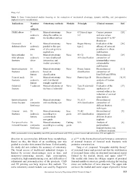
Table 2. Bone biomechanical studies focusing on the evaluation of mechanical advantage, dynamic stability, and post-operative
implants-relative complications.
Indications Number Osteotomy methods Module Principle Clinical measure Ref.
of
subjects
PMRI elbow 7 Manual osteotomy None O’Driscoll type Contact pressure [25]
cadaveric along the outline on 2-2 and area values
arms the trochlear surface throughout 0° to 90°
flexion arc
Coronoid- 8 Manual osteotomy None Regan-Morrey Evaluation of the [26]
deficient elbow cadaveric parallel to flat spot type 2 efficacy of coronoid
arms of ulna using micro prosthesis in elbow
sagittal saw stabilization
Intra-articular 16 Manual osteotomy None 41-C2 Biomechanical [24]
tibial plateau cadaveric in meta-diaphyseal AO-classification comparison of
fractures tibiae intersection and intramedullar versus
medial epicondyle extramedullar
stabilization
Intertrochanteric 16 Manual osteotomy None Evans-Jensen Biomechanical [11]
fractures cadaveric based on classification comparison of
femurs classification InterTAN and PFNA
Femoral neck 30 Manual osteotomy None Pauwel type B Biomechanical [4,10]
fractures cadaveric with thin bladed analysis of fixation
femurs straight sagittal saw devices
Odontoid 7 cadavers Manual osteotomy at None Type II odontoid Evaluation of the [27]
fracture the base of odontoid fracture application of
cervical collar in the
reduction of cervical
spine motion
Intertrochanteric 24 Manual osteotomy None 31-A3.3 Biomechanical [5]
femur fracture composite with oscillating saw AO-classification comparison of
Sawbones different fixation
techniques
Femoral neck 20 Manual osteotomy Saw- 31-B2 Biomechanical [9]
fractures cadaveric according to a guide AO-classification comparison of
femora custom-made different fixation
saw-guide implants
Periprosthetic 26 Manual osteotomy Cutting N/A Analysis of [8]
Femoral Fracture composite according to a cutting guide periprosthetic
Sawbones guide femoral fracture risk
in different implants
PMRI, posteromedial rotatory instability; InterTAN, proximal femoral nail; PFNA, proximal femoral nail anti-rotation; N/A: not applicable
direction to accommodate different bone morphology. et al. [12] in the biomechanical performance comparison
Based on this, the blade of an oscillating saw was of internal fixation of femoral neck fractures with
guided in circular slots around the bone. Unfortunately, posterior comminution.
the study did not evaluate the accuracy of osteotomy. Tang et al. proposed “triangular stability theory”
The broad access of traditional industrial designs is of proximal femur and pointed out that the stabilization
limited by several disadvantages such as time- and of proximal femur relies on structural mechanical model
manpower-consuming aspects, high consumption, formed by the medial, lateral, and upper sides [13,28] .
complexity, high manufacturing cost, and difficulties of The medial side forms the oblique support of proximal
refinement and personalization of the products. CAD femoral cantilever structure, greatly reducing the bending
and 3D printing can help improve this outlook. The stress and deflection of bone structure. The upper side
same osteotomy-aided module was used by Rupprecht connects the medial and lateral edges of proximal femur
International Journal of Bioprinting (2022)–Volume 8, Issue 4 113