
Page 184 - IJB-8-4
P. 184
A Review on Bioinks and their Application in Plant Bioprinting
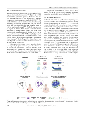
2.1. Scaffold-based bioinks At present, scaffold-based bioinks are the most
Scaffold-based bioinks can combine cells into an exogenous popular, due to their better innate structural properties,
.
scalability, reproducibility, and affordability
[37,38,42]
support structure for biomaterials [37-39] (Figure 2). dECM,
microcarriers (spherical, porous structures that aid 2.2. Scaffold-free bioinks
cell adhesion and growth), and hydrogels are common
components of this supporting scaffold (dECM) . The Scaffold-free bioinks are produced entirely using cells
[37]
scaffold further aids in the creation of functional tissue by and their generated matrices, thus they do not require any
promoting proliferation, differentiation, and cell growth additional biomaterials for support [37,38,40] . Scaffold-free
while also providing biological and chemical cues, as bioinks also refer to cells without the use of any exogenous
[38]
well as mechanical strength . Scaffolds are supposed biomaterial . Cell aggregation structures such as tissue
[40]
to degrade in time to form desired tissues while cells strands, cell sheets, pellets, and spheroids make up these
proliferate . Scaffold-based bioinks are often used bioinks, which rely on the capacity of cells to self-assemble
[40]
because their degradation rate is similar to the rate at into bigger tissue structures [37,38,41] . Scaffold-free bioinks
which cells construct the ECM . During the degradation waive the requirement for substantial cell growth due to
[41]
process, scaffold biomaterials disintegrate, allowing living tissue biomimicry, which improves cellular interactions,
cells to occupy the new space and form a predesigned high seeding densities, and reduces immunological
tissue structure . Controlling differentiation factors and responses in vivo [40-42] . Using scaffold-free bioinks, living
[3]
rate of growth are optimized when the breakdown rate of cells are printed in a manner that directly mimics normal
the scaffold can be regulated . embryonic growth . Clusters of the cells are deposited in
[3]
[41]
Although scaffold-based bioinks are often highly a specific pattern to build larger, integrated, and functional
biocompatible, they are sometimes prone to interruption tissue structures . Scaffold-free bioinks are promising
[37]
of cell-to-cell interactions, material toxicity, slow in tissue fabrication since they are biocompatible,
degradation rates, undesirable immune reactions during can facilitate ECM deposition with good cell-to-cell
in vivo testing, and compromised mechanical properties interaction, spread cells in a 3D environment, and enable
due to the complete deterioration of the scaffold [42-44] . the deposition of a high cell density . Bioink instigates
[45]
Figure 2. Diagrammatic illustration of scaffold-based and scaffold-free tissue engineering system. (from ref. licensed under Creative
[47]
Commons Attribution license). The figure was created with BioRender.com.
176 International Journal of Bioprinting (2022)–Volume 8, Issue 4