
Page 185 - IJB-8-4
P. 185
Ghosh and Yi
rapid differentiation and accelerates tissue maturation, speed, 600 mm min ) printing settings were optimized.
−1
preserves cell functions and features for a long period Bioink was cured with a built-in UV pen that followed the
of time, and provides a biomimicry microenvironment print path at a rate of 10 mm min . With the help of this
−1
to increase ECM production, thus minimizing the time bioink, a 0° – 90° grid was printed to analyze variations
required for adaptation to an external environment and in cell alignment across various z-planes . The cell
[48]
potentially overcoming rejection and tissue failure events culture was refilled 30 min after the support material was
that are common with scaffolds . removed to ensure that the cellular behavior observed in
[46]
Therefore, scaffold-free bioinks provide high the time point investigation of the bioprinted construct
resolution and cell viability, carefully mimic the cell was attributable to cells embedded within the hydrogel.
microenvironment of native organs and tissue for cell This prevents cells from dislodging from uncrosslinked
differentiation and proliferation, and preserve cell materials and adhering to the surface of struts. The printed
functionality and phenotypes for long periods of time . and seeded structures were cultured with cell media (high
[9]
glucose DMEM, 15% FBS, 1% Penstrep) at 37°C and
3. Bioinks and tissue engineering 5% carbon dioxide (CO ). The cell culture media were
2
[48]
3D bioprinting allows the precise geometrical control of replaced every 2 – 3 days .
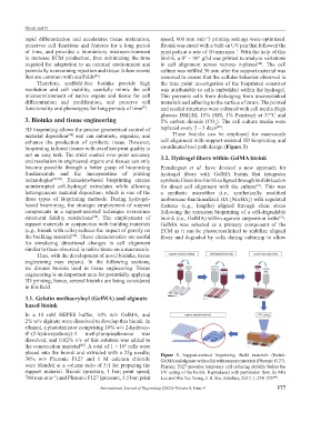
material deposition and can automate, organize, and These bioinks can be employed for macroscale
[48]
enhance the production of synthetic tissue. However, cell alignment with support-assisted 3D bioprinting and
bioprinting tailored tissues with excellent print quality is coordinated tool path design (Figure 3).
not an easy task. The strict control over print accuracy 3.2. Hydrogel fibers within GelMA bioink
and resolution in engineered organs and tissues can only
become possible through a better grasp of bioprinting Prendergast et al. have devised a new approach for
fundamentals and the incorporation of printing hydrogel fibers with GelMA bioink that integrates
technologies [49,50] . Extrusion-based bioprinting creates synthetic fibers into bioinks aligned through biofabrication
uninterrupted cell-hydrogel extrudates while allowing for direct cell alignment with the culture . This was
[51]
heterogeneous material deposition, which is one of the a synthetic microfiber (i.e., synthetically modified
three types of bioprinting methods. During hydrogel- norbornene-functionalized HA [NorHA]) with regulated
based bioprinting, the strategic employment of support features (e.g., lengths) aligned through shear stress
components in a support-assisted technique overcomes following the extrusion bioprinting of a cell-degradable
structural fidelity restrictions . The employment of bioink (i.e., GelMA) within agarose suspension baths .
[50]
[51]
support materials in conjunction with building materials GelMA was selected as a primary component of the
(e.g., bioink with cells) reduces the impact of gravity on ECM as it can be photocrosslinked to stabilize aligned
the building material . These characteristics are useful fibers and degraded by cells during culturing to allow
[48]
for simulating directional changes in cell alignment
similar to those observed in native tissue on a macroscale.
Thus, with the development of novel bioinks, tissue
engineering may expand. In the following sections,
we discuss bioinks used in tissue engineering. Tissue
engineering is an important area for potentially applying
3D printing; hence, several bioinks are being considered
in this field.
3.1. Gelatin methacryloyl (GelMA) and alginate
based bioink
In a 10 mM HEPES buffer, 10% w/v GelMA, and
2% w/v alginate were dissolved to develop this bioink. In
ethanol, a photoinitiator comprising 10% w/v 2-hydroxy-
4′-(2-hydroxyethoxy)-2 methylpropiophenone was
dissolved, and 0.02% v/v of this solution was added to
the construction material . A total of 1 × 10 cells were
[48]
6
placed into the bioink and extruded with a 25g needle; Figure 3. Support-assisted bioprinting. Build materials (bioink:
30% w/v Pluronic F127 and 1 M calcium chloride GelMA and alginate with cells) with support materials (Pluronic F127).
were blended at a volume ratio of 3:1 for preparing the Pluronic F127 provides temporary cell culturing stability before the
support material. Bioink (pressure, 1 bar; print speed, UV curing of the bioink. Reproduced with permission from Jia Min
700 mm min ) and Pluronic F127 (pressure, 3.5 bar; print Lee and Wai Yee Yeong, J. R. Soc. Interface, 2017, 1, 234–235 .
−1
[48]
International Journal of Bioprinting (2022)–Volume 8, Issue 4 177