
Page 263 - IJB-8-4
P. 263
Xu, et al.
nano- (1 nm to 1 μm) and micro-structural (1 μm to 1 mm) recreated from basic cells and materials. Complexity can
scale, cells proliferate, differentiate, and utilize their be also found at the interfaces between tissues, such as
inherent mechanisms to form nanostructures as organ the transition from cartilage to bone in the osteochondral
[10]
scaffolds. With the angiogenesis develops, a network of interface in articulating joints . An increase in the level
capillaries forms, providing cells with essential nutrients of complexity of the tissue or organ to be repaired usually
and growth factors. At the meso- (1 mm to 1 cm) and requires a corresponding increase in the complexity of
macrostructural (>1 cm) scale, identifiable tissues and the tissue engineering approach. 3D bioprinting offers
organs are formed from various types of cells, blood the best potential in deposition of biomaterials (with
vessels, extracellular matrix, etc. In general, there are or without proteins, growth factor, etc.,) and cells into
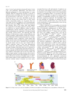
four scales (levels) of complexity in the macrostructure precise geometries to create anatomically correct living
of tissues and organs (Figure 1A). Flat tissues (relatively structures with multiscale.
thin structure, such as the cornea, skin, and bladder) Cellular responses to the surrounding environment
are composed of cell sheets stacked in multiple layers, are thought to play an important role in the pathogenesis
and allowing for nutrient diffusion without complete from developmental morphogenesis to disease states.
vascularization; Tubular structures (such as the artery, Cellular matrix elasticity can be used to facilitate
trachea, and urethra) are composed of cell sheets formed expected cellular behavior [11,12] . Human cells and tissues
into circular, bilayer tissues. The relatively thin wall have their own rigidity, ranging from a few kPa to
thickness of tubular tissue enables nutrient diffusion and tens of GPa (kidney [13,14] , heart [15,16] , skin [17,18] , arterial
[19]
[23]
oxygen exchange. Large-diameter vessels are relatively wall [19,20] , liver [21,22] , prostate , saphenous vein ,
easy to fabricate, while microstructural arterioles, venules, cornea [24,25] , breast [26,27] , tendon/ligament [28,29] , cancellous
and capillaries remain a challenge; Viscus organs with bone [19,30] , brain [31-33] , cartilage [34,35] , and cortical bone ),
[36]
hollow tubules (such as the heart, intestine, and stomach) depending on the specific cell type, extracellular matrix,
consist of an inner layer of epithelial cells surrounded and structural organization. Biomaterials that closely
by smooth muscle and an outer layer formed with or mimic organ nanostructures could be used to replicate
without connective tissue. It has been demonstrated nano-to-macro approach to human organ development,
that macroscopic and partially mesoscopic structural and proper biomaterial placement is necessary to direct
elements of organs with hollow tubes can be fabricated in cellular self-assembly [37,38] . The elastic modulus and
vitro . However, reconstructing organ microarchitecture, molding process characteristics of common printable
[8]
including glands and blood vessels, remains a challenge; biomaterials (collagen [39,40] , fibrin [41,42] , alginate ,
[43]
most complex solid organs (such as the liver, brain, and chitosan [44,45] , agarose , Poly(3-Hydroxybutyrate-co-3-
[46]
kidney) require mature vascular networks with extensive Hydroxyvalerate) (PHBV) , and other polymers [48,49] )
[47]
branching for cells to remain viable and function, as well as are summarized in Figure 1B. To print physically similar
precise distribution structures of multiple types of cells . microenvironments in vitro, appropriate mechanical
[9]
Solid organs require several essential structures to restore properties and modeling processes of the materials used
function, whereas tubular structures are more easily must be considered [50,51] . A highly biomimetic tissue/organ
A
B
Figure 1. (A) Organ anatomy by structural complexity. (B) Mechanical properties and molding process characteristics of biomaterials.
International Journal of Bioprinting (2022)–Volume 8, Issue 4 255