
Page 78 - IJB-8-4
P. 78
3D Printable PLA/BG Composite In Vitro Evaluation
A B A B
C D
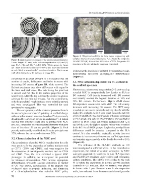
Figure 2. 3D-printed scaffolds for bone tissue engineering with
Figure 1. Light microscope images of the test specimens (diameter: complex internal structure made of pure PLA (A) and the composite
5 mm, height: 0.3 mm) with section magnification. (A) and (C) PLA/BG 20% (B). Even with a BG content of 20%, fine porous 3D
pure PLA, (B) and (D) PLA/BG 20%. White arrows in figure structures in the sub-millimeter range can be realized.
(B) show holes and dehiscence between the individual filament
strands. Black arrows show BG particles at interface. Black arrows evidencing the presence of sulfated glycosaminoglycans,
with white frame show BG particles in image (D). demonstrated successful chondrogenic differentiation
(Figure 3).
concentration at about 200 µm. It is noticeable that the
number of cracks, dehiscence, and holes increases with 3.3. MSC adhesion dependent on BG content in
increasing BG content (Figure 1B, white arrows). The the scaffold specimen
flat test specimens used show differences with regard to
the front and back sides. The side facing the print bed Fluorescence microscopy images taken 24 h post-seeding
is smooth and flat (due to the surface properties of the revealed MSC in comparatively low density on PLA as
heated bed), while the top side has the desired roughness BG material. Cell density increased with BG content
achieved by the printing process. For the experiments, and visually reached the highest densities at 10% and
only the populated rough surfaces were pointing upward 20% BG content. Furthermore, Figure 4E-H shows
and were investigated. This was controlled for each BG-dependent colonization with MSC. The cell density
specimen by a magnifier. increases with increasing BG content. The MTT assay
The core property of the material presented here is revealed an increase in metabolic activity of cells with the
its use in high-resolution 3D printing. A scaffold design higher BG contents. At 24 h time point, metabolic activity
with complex internal structure based on PLA previously of BG10 and BG20 was significantly enhanced compared
developed by our group served as a reference . It turned to PLA group, and cells in BG20 material showed higher
[1]
out that this structure could also be printed with PLA/ activity as BG5. These differences decreased after 72 h
BG 20% composite. Porous structural elements with pore and 168 h. Here, the highest median values were recorded
sizes of less than 150 µm are possible (Figure 2). Total in the BG10 and BG20 groups, despite that, no significant
porosity estimated by modified Archimedes principle was differences could be detected compared to the PLA
32%, whereas the calculated value was 28%. control. It is also noted that metabolic activity does not
continue to increase over time on any scaffold specimens
3.2. Characterization of the MSC pool and remains approximately at the levels detected at 24 h
Characterization of pooled MSCs revealed that the cells (Figure 4I).
were positive for the expression of surface markers such The influence of the PLA/BG scaffolds on OD
as CD73, CD90, and CD105, and were negative for was investigated at different levels. In the coincubation
the expression of hematopoietic markers such as CD34 experiment, the extent of calcium deposition by MSC
and CD45. Thus, the cells exhibited a typical MSC was semi-quantitatively analyzed in pure PLA specimen
phenotype. In addition, we investigated the potential and PLA/BG20 specimen under control and osteogenic
for trilineage differentiation. After running appropriate culture conditions. The MSCs were seeded to the well
differentiation protocols, calcium deposition as marker 48 h before the experiment in normal growth medium
of OD was shown, accumulation of fat droplets was (control) or OD medium. The test material was placed
evidenced after adipogenic induction, and formation in inserts with 3 µm diameter pores in the respective
of a dimethylmethylene blue stainable MSC pellet, wells, ions released from the test material could pass the
70 International Journal of Bioprinting (2022)–Volume 8, Issue 4