
Page 82 - IJB-8-4
P. 82
3D Printable PLA/BG Composite In Vitro Evaluation
A beneficial rheological properties structures with feature
sizes down to 100 µm, PLA can be readily fabricated
using commercially available 3D printers. Furthermore, it
is 100% biodegradable. Unfortunately, it has low affinity
for cells such as MSC or human osteosarcoma cell line
(Saos-2) and exerts no or only little osteoconductive or
osteoinductive influence on applied cells, such as MSC
or endothelial progenitor cells (EPCs) [1,7] . Instead, a
composite of PLA with BG showed a stimulation of EPC
differentiation and function. Apoptosis rate compared to
pure PLA was decreased [7,42] .
B For composite preparation, both components are
mixed and melted together. The BG component used here
is commercially available in various particle sizes. For
the filament used in this study, BG granules in the size
range of 25-42 µm were used. The integrated BG particles
require the application of a nozzle with a diameter of at
least 400 µm for printing to avoid clogging. Still, fine
structures with a resolution below to one-fourth of the
nozzle diameter could successfully be printed.
The material exhibits a homogeneous distribution of
C BG particles, which are localized both near the surface
and centrally in the filament (Figure 1). This allows
the continuous release of bioactive ions during the
degradation process. The dose-dependent accumulation
of calcium ions from the test particles in initially calcium-
free PBS could be demonstrated by us . The calcium
[37]
concentration was about 0.35 nmol/µL after 7 days of
incubation of a BG20 sample. Moreover, it is conceivable
that the BG particles alter roughness of the PLA surface,
which could facilitate the attachment of MSCs . This is
[43]
clearly indicated by the SEM images with the increased
cell affinity with increasing BG content (Figure 4).
The core property of the composite material
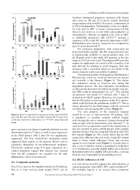
Figure 6. Relative expression of osteogenic marker genes RUNX2 should be its use in high-resolution 3D printing. This
(A), ALP (B), and COL1A (C) in MSC cultured 24, 72, and 168 h is mandatory to construct complex scaffold designs
on the test materials as indicated (n=5). *P<0.05 versus indicated with biologically active structural elements beyond the
group. classical grid with its monotonous pore structure [1,31] . In
addition to the test specimens used for the experiments, it
gene expression was almost completely inhibited over the was also possible to print scaffold designs with complex
observation period of 7 days, as well as gene expression internal structure from the composite filaments with 20%
of the MAP kinases JNK-1 and p38 was significantly BG content (Figure 2). This clearly shows that the BG
suppressed. The final whole-blood stimulation assays content also influences the print result. A high BG content
supported these results, as the material did not lead to leads to lower strand adherence. Pores and crevices
significant stimulation of pro-inflammatory mediators. appear on a micrometer scale (Figure 1). Overall, there is
also a loss of stability, which is quantified by Schätzlein
Mechanistic analyses using IL-6 gene expression as a et al. Furthermore, porosity slightly differs due to the fact
readout parameter suggest that calcium ions released that printed parts have slightly different surface, resulting
from the BG component are causative for the anti- from the layer topography by the printing process.
inflammatory effect of the material.
4.2. BG20: Influence on OD
4.1. Composite material
It is well known that BG supports the differentiation of
Pure PLA is used as the standard material in 3D printing. various progenitor cells (MSC, EPC, and osteoblasts) .
[44]
Due to its high melting point around 200°C and its For example, Aguirre et al. showed that a composite
74 International Journal of Bioprinting (2022)–Volume 8, Issue 4