
Page 80 - IJB-8-4
P. 80
3D Printable PLA/BG Composite In Vitro Evaluation
A B MSC calcium deposition after 14 days of incubation
under control conditions. Furthermore, BG20 did not
promote increase in calcium deposition under osteogenic
conditions compared to the PLA test specimen (Figure 5).
Next, the effect of the BG component on the
expression of prototypical osteogenic marker genes was
analyzed over time for 7 days. Gene expression of the
C D osteogenic transcription factor RUNX-2 was increased
after 24 h on median in the BG10 and BG20 groups
compared to PLA group, however, this was not statistically
significant after alpha correction was applied (p* = 0.01
BG10 vs. PLA, p* = 0.03 PLA vs. BG20). In the further
course, RUNX-2 gene expression in the BG10 and BG20
groups decreased, in some cases significantly, compared
to the corresponding 24 h values. ALP gene expression
E F
was initially increased in the BG5 group, however, it
was significantly elevated throughout time only in BG20
preparations. At time 168 h, ALP gene expression was
significantly increased in BG20 group compared to
BG10 group and in trend (p = 0.08) compared to BG5
and PLA preparations. COL1A gene expression increased
significantly over the course to 72 h in some cases (BG10
G H group) and declined to the 24 h levels after 168 h. No
significant differences on the relative expression of
COL1A could be detected among the different groups at
any time point (Figure 6).
3.4. Influence of BG scaffolds on inflammatory
processes
The regulation of inflammatory signaling pathways in
MSC, which are involved in cell differentiation processes
and play an important role in inflammatory response
in situ, was investigated at the gene expression level.
Results demonstrate that IL-6 gene expression was
significantly suppressed with increasing BG content
(BG10, BG20) over the entire observation period. The
MAP kinases (MAPK8 = JNK1, MAPK14 = p38) are
involved in signal transduction, most of which lead
to a pro-inflammatory response through activation of
NFkB. In particular, relative expression of MAPK8 was
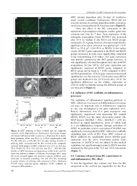
Figure 4. MSC adhering to PLA (control) and the composite significantly downregulated in MSC cultured on scaffolds
material. (A-D) Representative fluorescence microscope images containing high levels of BG. Thus, MSC cultured on
of Calcein-AM-stained MSC seeded on a 3D-printed PLA double BG20 exhibited the significantly lowest MAPK8 gene
layer specimen, either pure PLA (a) or PLA supplemented with expression over the entire observation period. Gene
5% (B), 10% (C), or 20% BG 24 h (D) after seeding. Number of
adhering MSC increased with the BG content in the composite expression of MAPK14 was not subject to this marked
material. SEM images of MSC cultivated over 24 h on PLA (E), regulation, but significant suppression by BG20 was also
PLA+5%BG (F), PLA+10% BG (G), and PLA + 20% BG (H). measured for this gene at the 72 h and 168 h time points
Density of MSC increased with percentage of BG in the scaffold. (Figure 7).
Scale bars indicate a distance of 200 µm. Accordingly, MTT assay
(E) (n=5) revealed higher cell metabolic activity, if MSCs were 3.5. Calcium released from BG contributes to
cultivated on material containing 10% and 20% BG, indicating anti-inflammatory BG effect
a greater number of adhering cells compared to PLA. *Indicates
P<0.05 versus indicated group. Influence of BG scaffolds on To test the hypothesis that calcium ions from the BG
osteogenic differentiation. component of the scaffold are responsible for the IL-6
72 International Journal of Bioprinting (2022)–Volume 8, Issue 4