
Page 84 - IJB-8-4
P. 84
3D Printable PLA/BG Composite In Vitro Evaluation
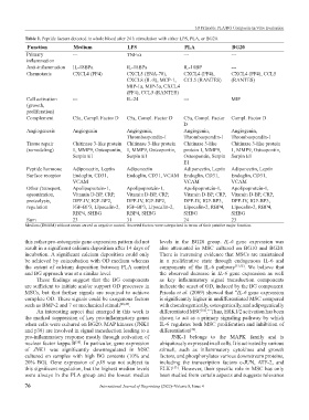
Table 1. Peptide factors detected in whole blood after 24 h stimulation with either LPS, PLA, or BG20.
Function Medium LPS PLA BG20
Primary --- TNF-α --- ---
inflammation
Anti-inflammation IL-18BPa IL-18BPa IL-18BP ---
Chemotaxis CXCL4 (PF4) CXCL5 (ENA-78), CXCL4 (PF4), CXCL4 (PF4), CCL5
CXCL8 (IL-8), MCP-1, CCL5 (RANTES) (RANTES)
MIP-1a, MIP-3a, CXCL4
(PF4), CCL5 (RANTES)
Cell activation --- IL-24 --- MIF
(growth,
proliferation)
Complement C5a, Compl. Factor D C5a, Compl. Factor D C5a, Compl. Factor Compl. Factor D
D
Angiogenesis Angiogenin Angiogenin, Angiogenin, Angiogenin,
Thrombospondin-1 Thrombospondin-1 Thrombospondin-1
Tissue repair Chitinase 3-like protein Chitinase 3-like protein Chitinase 3-like Chitinase 3-like protein
(remodeling) 1, MMP9, Osteopontin, 1, MMP9, Osteopontin, protein 1, MMP9, 1, MMP9, Osteopontin,
Serpin E1 Serpin E1 Osteopontin, Serpin Serpin E1
E1
Peptide hormone Adiponectin, Leptin Adiponectin Adiponectin, Leptin Adiponectin, Leptin
Surface receptor Endoglin, CD31, Endoglin, CD31, VCAM Endoglin, CD31, Endoglin, CD31,
VCAM VCAM VCAM
Other (transport, Apolipoprotein-1, Apolipoprotein-1, Apolipoprotein-1, Apolipoprotein-1,
opsonization, Vitamin D-BP, CRP, Vitamin D-BP, CRP, Vitamin D-BP, CRP, Vitamin D-BP, CRP,
proteolysis, DPP-IV, IGF-BP2, DPP-IV, IGF-BP2, DPP-IV, IGF-BP3, DPP-IV, IGF-BP3,
regulation IGF-BP3, Lipocalin-2, IGF-BP3, Lipocalin-2, Lipocalin-2, RBP4, Lipocalin-2, RBP4,
RBP4, SHBG RBP4, SHBG SHBG SHBG
Sum 23 31 24 23
Medium (DMEM) without serum served as negative control. Secreted factors were categorized in terms of their putative major function.
this rather pro-osteogenic gene expression pattern did not levels in the BG20 group. IL-6 gene expression was
result in a significant calcium deposition after 14 days of also attenuated in MSC cultured on BG10 and BG20.
incubation. A significant calcium deposition could only There is increasing evidence that MSCs are maintained
be achieved by coincubation with OD medium whereas in a proliferative state through endogenous IL-6 and
the extent of calcium deposition between PLA control components of the IL-6 pathway [51,52] . We believe that
and BG approach was at a similar level. the observed decrease in IL-6 gene expression as well
These findings suggest that the BG components as key inflammatory signal transduction components
are sufficient to initiate and/or support OD processes in indicate the onset of OD, induced by the BG component.
MSCs, but that further signals are required to achieve Pricola et al. (2009) showed that “IL-6 gene expression
complete OD. These signals could be exogenous factors is significantly higher in undifferentiated MSC compared
such as BMP-2 and 7 or mechanical stimuli [48,49] . with chondrogenically, osteogenically, and adipogenically
An interesting aspect that emerged in this work is differentiated MSC .” Thus, ERK1/2 activation has been
[52]
the marked suppression of key pro-inflammatory genes shown to act as a primary signaling pathway by which
when cells were cultured on BG20. MAP kinases (JNK1 IL-6 regulates both MSC proliferation and inhibition of
and p38) are involved in signal transduction leading to a differentiation .
[52]
pro-inflammatory response mostly through activation of JNK-1 belongs to the MAPK family and is
nuclear factor kappa B . In particular, gene expression ubiquitously expressed in all cells. It is activated by various
[50]
of JNK1 was significantly downregulated in MSC stimuli, such as inflammatory cytokines and growth
cultured on samples with high BG contents (10% and factors, and phosphorylates various downstream proteins,
20% BG). Gene expression of p38 was not subject to including the transcription factors c-JUN, ATF-2, and
this significant regulation, but the highest median levels ELK1 . However, their specific role in MSC has only
[53]
were always in the PLA group and the lowest median been studied from certain aspects and suggests relevance
76 International Journal of Bioprinting (2022)–Volume 8, Issue 4