
Page 79 - IJB-8-4
P. 79
Söhling, et al.
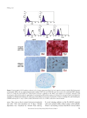
Figure 3. Representative FACS-analysis is shown in (A). Antigen expression (black line) and respective isotype controls (filled histograms)
are presented. MSCs did not express hematopoietic markers CD45 (A) and CD34 (B) and were positive for established MSC antigens
CD73 (C), CD90 (D), and CD105 (E). Differentiation potential is depicted in (B). MSCs were capable for osteogenic, adipogenic and
chondrogenic differentiation after the application of corresponding differentiation protocols. Evidence for osteogenic (calcium precipitation
visualized by Alizarin Red staining) (B), adipogenic (intracellular lipid droplets visualized by Oil Red O staining) (D), and chondrogenic
(DMMB staining after 21 days in pellet culture) differentiation (E). (A, C) MSCs cultured in control medium.
pores. There was no direct contact between test materials To avoid staining artifacts on the PLA/BG20 material,
and cells. Cells were incubated for 14 days. Calcium cells were cultured without direct contact with scaffolds.
deposition was visualized by Alizarin Red staining. Alizarin red staining revealed that BG20 did not induce
International Journal of Bioprinting (2022)–Volume 8, Issue 4 71