
Page 286 - IJB-9-1
P. 286
International Journal of Bioprinting 3D-Printed scaffolds
A B C
Figure 1. Fourier transform infrared of poly(ε-caprolactone) (PCL)/β-tricalcium phosphate (TCP) (A), poly(trimethyl carbonate) (PTMC)/TCP (B), and
PTMC/PCL/TCP (C) scaffolds with different TCP content.
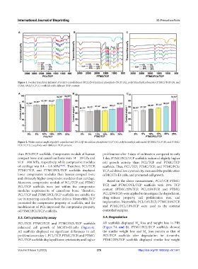
A B C
Figure 2. Water contact angle of poly(ε-caprolactone) (PCL)/β-tricalcium phosphate (TCP) (A), poly(trimethyl carbonate) (PTMC)/TCP (B), and PTMC/
PCL/TCP (C) scaffolds with different TCP content.
than PCL/TCP scaffolds. Compressive moduli of human proliferation after 3 days of cultivation compared to only
compact bone and cancellous bone were 14 – 20 GPa and 1 day. PTMC/PCL/TCP scaffolds indicated slightly higher
97.8 – 800 MPa, respectively, while compressive modulus cell growth activity than PCL/TCP and PTMC/TCP
of cartilage was 0.4 – 0.8 MPa [32,33] . Therefore, PCL/TCP, scaffolds. Thus, PCL/TCP, PTMC/TCP, and PTMC/PCL/
PTMC/TCP, and PTMC/PCL/TCP scaffolds displayed TCP exhibited low cytotoxicity, increased the proliferation
lower compressive modulus than human compact bone of MC3T3-E1 cells, and promoted cell growth.
and obviously higher compressive modulus than cartilage.
Moreover, compressive moduli of PCL/TCP and PTMC/ Based on the above measurement, PCL/TCP, PTMC/
PCL/TCP scaffolds were just within the compressive TCP, and PTMC/PCL/TCP scaffolds with 25% TCP
modulus requirements of cancellous bone. Therefore, content (PTMC/25%TCP, PCL/25%TCP, and PTMC/
PCL/TCP and PTMC/PCL/TCP scaffolds are suitable for PCL/25%TCP) were applied to investigate the degradation,
use in repairing cancellous bone defects. Meanwhile, TCP drug -release property, cell proliferation rate, and
promoted the compressive property of scaffolds, and the implantation. Meanwhile, PCL/10%TCP, PTMC/10%TCP,
modification of PCL improved the compressive property and PTMC/PCL/10%TCP were used as the contrast
of PTMC/PCL/TCP scaffolds. controlled samples.
3.3. Cell cytotoxicity assay 3.4. Degradation
PCL/TCP, PTMC/TCP, and PTMC/PCL/TCP scaffolds All scaffolds displayed M loss and weight loss in PBS
n
enhanced cell growth of MC3T3-E1 cells (Figure 6). (Figure 7A and B). PTMC/PCL/TCP scaffolds showed
All scaffolds displayed no significant differences in cell the similar weight loss and M loss pattern as that of
n
proliferation on day 1. PCL/TCP, PTMC/TCP, and PTMC/ PCL/TCP scaffolds after degradation for 6 months.
PCL/TCP scaffolds displayed lower cytotoxicity and higher PTMC/25%TCP scaffolds displayed similar low weight
Volume 9 Issue 1 (2023) 278 https://doi.org/10.18063/ijb.v9i1.641