
Page 31 - IJB-9-1
P. 31
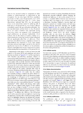
International Journal of Bioprinting                              Biocompatible materials and Multi Jet Fusion


            those  on the  uncoated surface in  comparison to  other   appeared and survived on seeding. Nonetheless, the electron
            coating or plasma-treatment, as determined at day   micrographs provided additional evidence showing the
            4 timepoint. On  the other  hand,  MC3T3e1 osteoblasts   presence and adherence of cells on the printed PA-12. In
            grew better on PDL-coated 3D-printed PA-12 (P < 0.05)   contrast, a completely different behavior was observed for
            and CLG-coated polystyrene plate (P < 0.05). These   osteoblasts which were bigger in size (>50 µm) compared
            observations indicated that PDL was the preferred   to  fibroblasts.  Osteoblasts  exhibited  a  flattened  shape  and
            choice as an extracellular matrix for culturing cells of   appeared to spread over multiple particles and had many
            both types on 3D-printed PA-12, even though it did not   prominent  filopodia  protrusions  stretching  over  multiple
            substantially narrow the gap with polystyrene plate. The   particles (Figure 4B  i–iii). As a result, the main cell body
            fact that fibroblasts survived better in the 3D-printed   appeared loosely attached and suspended between particles,
            PA-12 cell culture chambers than osteoblasts during   making it difficult for the osteoblasts to completely attach
            short-term culture was apparent. Cells demonstrated   and proliferate. Control PA-12 cell culture chambers
            equal  proliferation  on  uncoated  hydrophobic  PA-12   (medium only) are also shown for reference (Figure
            surfaces,  even  in  the  absence  of  surface  coating or  O    S11A i–iii  and  S11B i–iii). Fibroblast and osteoblasts cells
                                                          2
            plasma-treatment, indicating that the surface wettability   (and control samples without cells) grown on polystyrene cell
            did not significantly affect the cell adherence. When   culture chambers are also shown for comparison (Figure S11C
            substrates were plasma-treated, a significant decrease in   i–iii and S11D i–iii). Taken together, these findings supported
            hydrophobicity  was  observed  (P <  0.05). Even  though   the fact that 3D-printed PA-12 can support cell growth, but not
            substrates demonstrated higher wettability, this did not   to the extent observed on polystyrene plates, probably due to
            significantly improve the cell adhesion on the substrates.   the microroughness resulting from partially fused particles on
            Therefore, hydrophobicity was not considered a key factor   the surface among other factors.
            in determining the biocompatibility of 3D-printed PA-12.
                                                               3.3.3. Cellular toxicity
              Representative SEM images of L929 and MC3T3e1 cells
            cultured on 3D-printed PA-12 and polystyrene cell culture   The short-term exposure of L929 fibroblasts and MC3T3e1
            chambers are shown in Figure 4A–D. In all the experiments,   osteoblasts to uncoated, PDL- and CLG-coated 3D-printed
            L929 fibroblasts and MC3T3e1 osteoblasts cultured on   PA-12 cell culture chambers, and polystyrene plate resulted in
            coated and uncoated 3D-printed PA-12 cell culture chambers   minimal LDH secretion due to cellular membrane integrity
            presented a flat morphology and appeared intimately adherent   on day 2 and day 4, respectively. Consequently, cells cultured
            to the surface, with no specific orientation. L929 fibroblasts   on both coated (PDL and CLG) and uncoated 3D-printed
            and MC3T3e1 osteoblasts cultured on coated and uncoated   PA-12 demonstrated no signs of cytotoxicity. No significant
            3D-printed PA-12 cell culture chambers appeared intimately   difference in cytotoxicity was observed between coated and
            adherent to the surface and showed an increase in the number   uncoated substrates, as shown in Figure 5A and B.
            of cells attached to the surface, noticeable after 4 days of   3.3.4. Redox characterization
            incubation. Fibroblasts exhibited cellular dimensions (~15 µm
            in diameter)  (Figure 4A  i–iii) smaller than the unfused   Visualization of oxidative stress assessed by mBCI staining
            PA-12 particles, making it conducive for cells to adhere to a   of L929 fibroblasts and MC3T3e1 osteoblasts cultured on
            single particle. Interestingly, unlike in the fluorescent images,   3D-printed PA-12 cell culture chambers and polystyrene
            the morphology of L929 grown on PA-12 and polystyrene   plate showed preservation of intracellular reduced GSH on
            appeared different in the SEM images. Such differences could   both day 2 and day 4, regardless of the type of surface coating
            be devoted to intrinsic artifacts such as specimen shrinkage    (uncoated  and coated)  (Figure 5C  and  Figure S12).  For
                                                        [55]
            more likely as an effect of glyceraldehyde fixation  or ethanol   fibroblasts, GSH-mBCl fluorescence signal was comparable
                                                [56]
            dehydration   before  critical  point  drying.  Some  earlier   between cells grown on 3D-printed PA-12 cell culture
                     [57]
            studies had also suggested great variation of these artifacts   chambers and polystyrene plates at both time points,
            from one type of sample to another . It was also indicated   suggesting that cells were not significantly stressed on
                                        [58]
            that the use of high-resolution SEM had known to accentuate   culture in 3D-printed PA-12 cell culture chambers. However,
            artifacts which might otherwise appear minimal at a lower   osteoblasts cultured on 3D-printed PA-12 showed a reduction
            resolution . In addition, the influence of the MJF-printed   in the overall intensity when compared to that observed on
                   [59]
            PA-12’s surface properties (such as roughness) on preserving   polystyrene plate, possibly as a result of the lower cell count.
            cell appearance from the effect of SEM-processing methods
            had to be highlighted. Moreover, the literature had minimal   3.3.5. Cellular proliferation
            evidence on the effect of critical point drying on 3D-printed   The proliferative state of cells characterized by
            PA-12, which had a major role in deciding on how the cells   immunostaining of Ki67 and p53 suggested that


            Volume 9 Issue 1 (2023)                         23                      https://doi.org/10.18063/ijb.v9i1.623