
Page 34 - IJB-9-1
P. 34
Biocompatible materials and Multi Jet Fusion
A
B
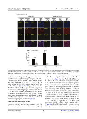
Figure 6. (A) Representative fluorescence microscopy images of L929 fibroblasts and MC3T3e1 osteoblasts immunostained to determine the expression of
ki67 and p53 following culture on PDL-coated 3D-printed PA-12 and positive control (plate) cell culture chambers after 4 days. (B) Alkaline phosphatase
activity (normalized) of MC3T3e1 cultured on uncoated, PDL- and CLG-coated 3D-printed PA-12 cell culture chambers at day 28.
of abnormality or changes in cell appearance, comparable uniformly covering the entire surface after 24 h,
to that observed on polystyrene plate (Figures S20–S25). as shown in Figure 8A. The bacterial viability on
The fibroblasts continued to grow robustly and proliferated 3D-printed PA-12 cell culture chambers was comparable
well when subcultured from PA-12 to PA-12 as well as to that observed on conventional culture plates (positive
from PA-12 to the polystyrene control plate as confirmed control), suggesting strong bacterial attachment. The big
by the MTT assay (Figure 7B and C). As observed in the green spheres in the background were due to the non-
short-term culture, fibroblasts grew better in comparison specific staining of the partially fused PA-12 particles.
to osteoblasts. After first passage, osteoblasts adhered to The contribution of red fluorescence, which represented
PA-12 cell culture chambers; however, a reduction in their
proliferation was represented by the decreased MTT signals the dead bacteria, was less predominant, indicating
which can be noted even in positive control at day 12. After that the fraction of dead bacteria was significantly
second passage, significant exhaustion of the MC3T3e1 lower. The viability experiments by MTT assay further
proliferative capacity was observed (Figure 7C). supported these results. Adhered bacteria on the surface
of PA-12 cell culture chambers (after 24 h), when tested
3.3.8. Bacterial viability and fouling directly for viability, indicated good bacterial activity
Examination of 3D-printed PA-12 cell culture chambers (Figure 8B). No inhibiting effect of PA-12 on the growth,
showed heavy and dense growth of bacteria (green) fouling, and viability of the bacterial cells was observed.
Volume 9 Issue 1 (2023) 26 https://doi.org/10.18063/ijb.v9i1.623