
Page 37 - IJB-9-1
P. 37
International Journal of Bioprinting Biocompatible materials and Multi Jet Fusion
protein binding capability using BSA-FITC. Earlier studies Both fibroblast and osteoblast cells show the ability to
demonstrate the use of low-pressure O plasma to activate grow in the leachate medium (Figure 2A and Figure S2).
2
porous surfaces for significantly increasing the utility Fibroblasts grow well regardless of whether it is cultivated
value of 3D-printed products by improving previously in the leachate or the normal medium. However,
inaccessible poor surface properties of the products . osteoblasts grown in the leachate demonstrate a drop in the
[64]
The protein adsorption experiments using BSA-FITC MTT values at day 4 compared to those grown in αMEM,
indicated successful dose-dependent protein capture that suggesting that they proliferate better in the normal
plateaus, suggesting saturation, at around 1%, indicating medium in comparison to the leachate (Figure 2B and 2C).
a possibility that the surfaces of 3D-printed materials can Although it is statistically different, the difference is small.
simulate the properties of natural extracellular matrix As aforementioned, the leachate medium is prepared by
aiming to regulate the behavior of cell adhesion (Table 1). soaking PA-12 cell culture chambers in the respective cell
This may also suggest that the spatial conformation of the culture media. Small molecules that leach into the medium,
adsorbed biomolecules plays a key factor in mediating possibly from fusing and detailing agents, may have caused
cell adhesion rather than the concentration or amount of this effect, as indicated in Table 1. Therefore, the growth
molecules affecting the adhesion process . However, there of a certain cell type is slower in leachate compared to the
[65]
is no significant difference between O plasma-treated and control. Otherwise, no other potential material leaching
2
untreated cell culture chambers (Figure 1G and H). In this issue is observed in our experiments as the typical test only
case, however, O plasma-treatment does not enhance uses leachate medium exposed to MJF-printed PA-12 for
2
the protein binding capability of MJF-printed PA12 . 5 days. Further, investigations are underway to unravel
[61]
Based on this observation, 3D-printed PA-12 are directly the effect of material leaching with regards to cell growth.
functionalized with biopolymers such as PDL and CLG However, further studies are required to probe into cell
(at 50 µg/mL), commonly used biomolecules that help in line specific in vitro toxicity and cytocompatibility of
initiating cell attachment and maintaining cell growth. 3D-printed PA-12.
The primary focus of this work is to evaluate the As shown in MTT studies from direct culture in
biocompatibility of 3D-printed PA-12 cell culture chambers. 3D-printed PA-12 cell culture chambers, L929 fibroblasts
The choice of assays plays a key role in assessing the material adapt well and proliferate efficiently when cultured in the
cytotoxicity, while other parameters, such as controls, cell 3D-printed PA-12 (Figure 3B and 7B) cell culture chambers,
lines, period of culture, and other biochemical events are whereas osteoblasts did grow and proliferate, but to a lesser
crucial in testing a material’s compatibility . Here, we study degree (Figure 3C and 7C). The observed reduction in
[66]
the biocompatibility of 3D-printed PA-12 by 2 methods: (i) proliferation of osteoblasts could be related to the fact that
by indirectly exposing the cells to the leachate medium that MTT activity is directly proportional to the cell number .
[67]
is extracted by exposing the culture medium to 3D-printed Moreover, osteoblasts appear larger in size compared to
PA-12 parts and (ii) by directly seeding the cells on the fibroblasts, as shown in Figure 3A and Figures S3–S10.
surface of the material (short- and long-term culture). This Many osteoblast cells (Figure 4B) are suspended between
study, which employs multiple cell lines, including L929 two particles, whereas fibroblasts fully attach on one
fibroblasts and MC3T3e1 osteoblasts grown on surface- particle. SEM images after 4 days of culture show that the
coated 3D-printed PA-12, shows a significant difference in L929 cells are more or less rounded in shape, orientate
their sensitivity toward the cell culture chambers. symmetrically with small cellular extensions aiding in
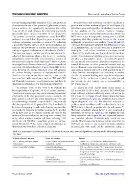
Table 1. A summary of key MJF‑printed PA‑12 features of interest, their associated problems, and our results addressing them in
this study
Printed substrate’s features Potential problems Results
PA-12’s binding affinity for protein • Ease of surface functionalization with • Printed PA‑12 can bind to BSA, indicating surface
biomolecules extracellular matrix modification with protein extracellular matrix is possible
Surface topography/roughness • Impaired cell adhesion • Cells can adhere to printed PA‑12 from passage to passage
• Abnormal cell morphology • While fluorescence microscopy does not indicate an obvious
change in cell morphology, SEM shows fibroblast displaying
unusual morphology
Fusing and detailing agents • Cytotoxicity • No cytotoxicity is detected
• Growth inhibition • Cells can proliferate, though not as good as on polystyrene
• Differentiation inhibition surface
• Osteoblast can differentiate
Volume 9 Issue 1 (2023) 29 https://doi.org/10.18063/ijb.v9i1.623