
Page 134 - IJB-9-2
P. 134
International Journal of Bioprinting Design and manufacture of high-performance bone plate
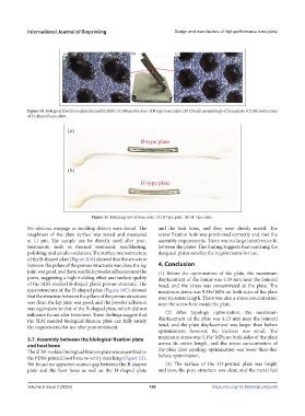
Figure 10. Biological fixed bone plate formed by SLM. (A) Microstructure of B-type bone plate. (B) Overall morphology of bone plate. (C) Microstructure
of H-shaped bone plate.
Figure 11. Matching test of bone plate. (A) B-type plate. (B) H- type plate.
No obvious warpage or molding defects were found. The and the host bone, and they were closely mated. The
roughness of the plate surface was tested and measured screw fixation hole was positioned correctly and met the
at 11 µm. The sample can be directly used after post- assembly requirements. There was no large interference fit
treatments, such as thermal treatment, sandblasting, between the plates. This finding suggests that matching the
polishing, and anodic oxidation. The surface microstructure designed plates satisfies the requirements for use.
of the B-shaped plate (Figure 10A) showed that the structure
between the pillars of the porous structures was clear, the lap 4. Conclusion
joint was good, and there was little powder adhesion near the (1) Before the optimization of the plate, the maximum
pores, suggesting a high molding effect and surface quality displacement of the femur was 4.09 mm near the femoral
of the SLM-molded B-shaped plate’s porous structure. The head, and the stress was concentrated at the plate. The
microstructure of the H-shaped plate (Figure 10C) showed maximum stress was 9.38e MPa on both sides of the plate
2
that the structure between the pillars of the porous structures over its entire length. There was also a stress concentration
was clear, the lap joint was good, and the powder adhesion near the screw hole inside the plate.
was equivalent to that of the B-shaped plate, which did not
influence its use after treatment. These findings suggest that (2) After topology optimization, the maximum
the SLM-molded biological fixation plate can fully satisfy displacement of the plate was 4.13 mm near the femoral
the requirements for use after post-treatment. head, and the plate displacement was larger than before
optimization; however, the increase was small. The
2
3.7. Assembly between the biological fixation plate maximum stress was 5.15e MPa on both sides of the plate
and host bone across its entire length, and the stress concentration of
The SLM-molded biological fixation plate was assembled to the plate after topology optimization was lower than that
the FDM-printed host bone to verify matching (Figure 11). before optimization.
We found no apparent contact gap between the B-shaped (3) The surface of the 3D-printed plate was bright
plate and the host bone as well as the H-shaped plate and new, the pore structure was clear, and the metal feel
Volume 9 Issue 2 (2023) 126 https://doi.org/10.18063/ijb.v9i2.658