
Page 154 - IJB-9-2
P. 154
International Journal of Bioprinting Enhanced osteogenesis in gelatin releasing bioink
(GAPDH); 5’-GCCAATCCCTAAGTGTGGCT-3’ (sense) and washed with PBS. Light microscopic images were
and 5’-ACATAGGTCCCCATCTGCCT-3’ (anti-sense) for taken with an optical microscope (IX71; Olympus, Japan)
runt-related transcription factor 2 (RUNX2); at a magnification of 10×.
5’-CCAGCCGCAAAGAGTCTACA-3’ (sense) and
5’-CTTGGGTCCCTCGACTCCTA-3’ (anti-sense) for 2.12. Statistical analysis
collagen type I (COL1); 5’-CAGGCCGCCTTCATAAGCA-3’ Data of water content, mechanical property, ALP activity,
(sense) and 5’-GTGCCGATGGCCAGTACTAA-3’ (anti-sense) and qPCR were analyzed by one-way analysis of variance
for ALP; 5’-GTTTGGCTTTAGGGCAGCAC-3’ (sense) and (ANOVA). Tukey simultaneous tests were used to identify
5’-GGGCAGCACAGGTCCTAAAT-3’ (anti-sense) for differences between individual hydrogel groups using
osteocalcin (OCN); and 5’-CCTTGCTTGGGTTTGCAGTC-3’ Origin software. Each cell activity experiment was repeated
(sense) and 5’-TTCTGTGGCGCAAGGAGATT-3’ (anti- at least three times with four different samples in each
sense) for osteopontin (OPN). group, and a P-value < 0.05 was considered statistically
significant.
2.10. Fabrication of cell-laden MA-alginate/gelatin
scaffold 3. Results and discussion
The cell-laden MA-alginate/gelatin mixture solution (at
either a 7:3 or 5:5 ratio of MA-alginate/gelatin) was stored Each MA-alginate/gelatin mixture solution (at MA-
at 4°C for 10 min. Then, the scaffold was fabricated using alginate/gelatin ratios of 10:0, 9:1, 7:3, and 5:5) was stored
a customized three-axis material extrusion-based printer at 4°C for 10 min before hydrogel disk fabrication to
with a nozzle (inner diameter = 390 μm) at the following allow the gelatin to solidify and become trapped in the
temperatures: syringe temperature of 15°C and plate MA-alginate. Regardless of the amount of gelatin in each
temperature of 10°C. The applied pneumatic pressure was hydrogel sample, at 37°C, MA-alginate transforms into a
80–100 kPa. The feed rate of the printing system was set at liquid phase, which is also the cell culturing temperature.
200 mm·min . Given that the system was designed to release gelatin
-1
from the matrix, we investigated the differences in the
2.11. Alizarin Red S staining characteristics of the hydrogel samples and their effects on
After 3 weeks of culture, the cell-laden scaffolds cells external to and encapsulated in the hydrogel samples.
containing different ratios of components (7:3 or 5:5
MA-alginate/gelatin) were fixed in 4% formaldehyde, 3.1. Characterization of MA-alginate/gelatin
dehydrated in a graded series of ethanol, and embedded hydrogel
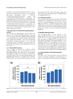
in paraffin blocks. After that, the sectioned samples were The water content was measured to assess the swelling
deparaffinized, hydrated in a graded series of ethanol, property of each type of hydrogel, as indicated in Figure 2a
stained with 2% (w/v) Alizarin Red S solution for 5 min, and 2b. It was measured at two time points: immediately
Figure 2. Water contents of hydrogels according to different volume ratio of MA-alginate/gelatin at (a) 0 h and (b) 3 weeks. *P < 0.05, n.s. = not significant.
Volume 9 Issue 2 (2023) 146 https://doi.org/10.18063/ijb.v9i2.660