
Page 158 - IJB-9-2
P. 158
International Journal of Bioprinting Enhanced osteogenesis in gelatin releasing bioink
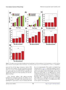
Figure 5. Cell activity of external cell affected by gelatin released from hydrogel. (a) DNA quantification. (b) ALP quantification. (c–g) qPCR results for
genes monitored in each group after 3 weeks of culturing (n = 3): (c) RUNX2, (d) COL1, (e) ALP, (f) OCN, and (g) OPN; *P < 0.05, n.s. = not significant.
genes for the mid-late stage osteogenesis markers OPN due to its retained gelatin. This could be concluded along
and OCN were evaluated. The gene expression associated with the results of Figure 5. From the osteogenic behavior
with the 5:5 and 7:3 MA-alginate/gelatin hydrogels did results of external cells and encapsulated cells, both released
not significantly differ; however, the gene expression was and retained gelatin, which was in higher amounts, had
significantly different from that associated with the MA- identically positive effect on cell differentiation. Gelatin
alginate hydrogel. contains the arginine-glycine-aspartic (RGD) sequence and
promotes cell adhesion and proliferation of connection-
These findings indicate that gelatin-containing dependent cells [32,33] . These improvements of cell adhesion
hydrogels have osteogenic potential. In addition, the 5:5 and proliferation in hydrogels and external cells might
MA-alginate/gelatin hydrogel has the highest potential for have a more prominent effect in cell differentiation than
inducing bone regeneration activity on encapsulated cells the non-gelatin-containing group.
Volume 9 Issue 2 (2023) 150 https://doi.org/10.18063/ijb.v9i2.660