
Page 162 - IJB-9-2
P. 162
International Journal of Bioprinting Enhanced osteogenesis in gelatin releasing bioink
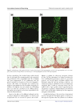
Figure 9. Live/dead assay on fabricated scaffold after 3 weeks of culturing: (a) 7:3 MA-alginate/gelatin and (b) 5:5 MA-alginate/gelatin, and in vitro osteo-
genic cellular activities through Alizarin red S staining: (c) 7:3 MA-alginate/gelatin and (d) 5:5 MA-alginate/gelatin.
for bone remodeling. The live/dead assay results showed alginate to gelatin for enhancing osteogenic behavior
that the hydrogels with remaining gelatin also supported in vitro. We then determined the optimal conditions for
the proliferation of the encapsulated cells. We concluded three scaffold printing parameters: nozzle size, pneumatic
that these enhancements of cell proliferation and cell pressure, and moving speed. Cell-laden scaffolds were
growth resulted from the presence of the remaining gelatin fabricated using the optimal printing conditions and
because cells within the hydrogels that contained less cultured in osteogenic media for 3 weeks. Live/dead assays
gelatin or were gelatin-free did not exhibit cell growth. In conducted after 3 weeks of culturing showed that the
addition, MA-alginate does not contain any cell-binding scaffold with a higher gelatin content supported greater
sites, so it does not contribute to the cellular activity, cell growth. In addition, calcium deposits were detected
leaving the retained gelatin as the factor responsible for the within both scaffold types, indicating that they supported
observed cell proliferation. osteogenic differentiation.
Based on the effects of the different hydrogels and the Overall, the findings of this study have demonstrated
associated released and retained gelatin on external and that the hydrogel with the highest gelatin content (a 5:5
encapsulated cells, we selected the optimal ratio of MA- MA-alginate to gelatin ratio) has potential as an effective
Volume 9 Issue 2 (2023) 154 https://doi.org/10.18063/ijb.v9i2.660