
Page 361 - IJB-9-2
P. 361
International Journal of Bioprinting Laser bioprinting of hiPSC-derived neural stem cells and neurons
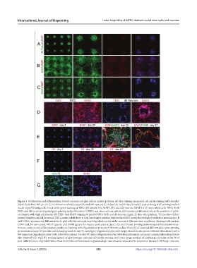
Figure 3. Proliferation and differentiation toward neurons and glial cells in printed patterns. All blue staining are general cell nuclei staining with Hoechst
33342. Scale bars: 100 µm. (A–C) Proliferation of NSCs (A and B) and d20 neurons (C) 2 days (A) and 12 days (B and C) post-printing. Ki67 staining marked
nuclei of proliferating cells in red, while green staining of NSCs (β3-tubulin (A); MAP2 (B)) and d20 neurons (MAP2 (C)) stains whole cells. While both
NSCs and d20 neurons migrated post-printing and proliferation of NSCs was observed everywhere, d20 neurons proliferated only at the positions of print-
ed droplets with high cell density. (D) TBR1 and MAP2 staining of printed NSCs (left) and d5 neurons (right) 22 days after printing. The position of four
printed droplets can still be seen in TBR1 panels, while there is only one droplet position depicted in MAP2 panels due to higher resolution microscopy. (E
and F) NSC spontaneous differentiation to glial cells like astrocytes and oligodendrocytes under neuronal differentiation conditions. Staining with markers
GFAP (red, for astrocytes), MAP2 (green), and S100B (green, for mature astrocytes) at days 2, 23, and 37 post-printing demonstrates differentiation to as-
trocytes under neural differentiation conditions. Staining with oligodendrocytic marker O4 (red) at days 35 and 67 of neuronal differentiation post-printing
demonstrated many O4-positive cells already present on day 35, mostly pre-oligodendrocytes with simple dendritic extensions (without bifurcations) and a
few immature oligodendrocytes (with a few bifurcations). On day 67, mature oligodendrocytes (with long extensions and many complex bifurcations) were
also observed. (G) vGLUT1 staining (green) of glutamatergic neurons and GABA staining (red, same image section) of GABAergic neurons at day 30 of
post-differentiation of printed NSCs. Most of the NSCs differentiated to glutamatergic neurons and only a smaller proportion became GABAergic neurons.
Volume 9 Issue 2 (2023) 353 https://doi.org/10.18063/ijb.v9i2.672