
Page 365 - IJB-9-2
P. 365
International Journal of Bioprinting Laser bioprinting of hiPSC-derived neural stem cells and neurons
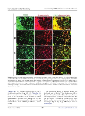
Figure 5. Formation of neuronal networks and synapses. Development of pre- and post-synaptic compartments by printed NSCs (A and B) and d20 neu-
rons (C and D), both neuronal post-differentiated for 20 days and stained with pre-synaptic synaptophysin (red) and post-synaptic density protein PSD95
(green) together with Hoechst 33342 cell nuclei staining (blue). (B and D) Details of (A) and (C), respectively. For each panel (A–D), the right image is a
merger of the others; the yellow color in these merged images is an overlap of red and green color. Both synaptic markers were found in abundance after 20
days of neuronal differentiation, and synaptic compartments could be seen in juxtaposition (red and green spot next to each other) and in colocalization
(yellow spot as overlap of red and green spot). This demonstrates neuronal maturation, extensive formation of synapses, and connection of neurons via
synapses. Scale bar: 50 µm.
Videoclip S2), with bursting events emerging by day 37 The spontaneous activity of neurons printed with
of differentiation (day 39 pp, diff 0/37; Videoclip S3). hyaluronic acid on Matrigel did also increase with the
TM
Further cultivation under differentiation conditions up duration of pre-differentiation (data not shown). However,
to day 65 of differentiation did not increase the average the average observed activity was always a bit lower than
observed activity. It must be noted, however, that there was that of NSCs and the number of average bursting events
always large variation across samples, with ones depicting directly visible was considerably lower. An illustrative
low activity and others exhibiting abundant and intense recording at day 26 (day 26 pp, diff20/26) is shown in
activity. Videoclip S4.
Volume 9 Issue 2 (2023) 357 https://doi.org/10.18063/ijb.v9i2.672