
Page 363 - IJB-9-2
P. 363
International Journal of Bioprinting Laser bioprinting of hiPSC-derived neural stem cells and neurons
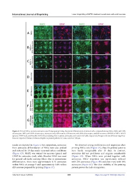
Figure 4. Printed NSCs, neurons and astrocytes 23 days post-printing. Neuronal differentiation of printed cells, compared among NSCs, NSCs with 20%
of astrocytes, NSCs with 50% of astrocytes, astrocytes only, d20 neurons, d20 neurons with 20% of astrocytes, and d20 neurons with 20% of NSCs. MAP2
(green), GFAP (red), and Hoechst 33342 (blue) staining show neurons, astrocytes, and nuclei of all cells, respectively. Images with two different magnifica-
tions are depicted. Distance between droplets in printed patterns is 1 mm; scale bar: 500 µm.
results are depicted in Figure 4. For comparison, astrocytes We observed strong proliferation and migration when
from astrocytic differentiation of NSCs were also printed printing NSCs only (Figure 4A), thus the printed patterns
and cultured for 23 days under neuronal culture conditions were barely recognizable after 23 days. In contrast,
(Figure 4D). MAP2 was stained for neurons (green) and astrocytes did not proliferate and migrate considerably
GFAP for astrocytes (red), while Hoechst 33342 was used (Figure 4D). When NSCs were printed together with
for general cell nuclei staining (blue). Due to spontaneous astrocytes, NSCs’ migration was significantly reduced
differentiation, there were approximately 0.1% astrocytes with 20% astrocytes (Figure 4B) and even more with 50%
within NSCs at passage 5 and approximately 0.4% within astrocytes (Figure 4C). The clear visibility of the printing
d20 neurons prepared for printing (Figure 1C). pattern proves the lack of migration.
Volume 9 Issue 2 (2023) 355 https://doi.org/10.18063/ijb.v9i2.672