
Page 466 - IJB-9-2
P. 466
International Journal of Bioprinting Bioprinting of exosomes
In a different approach, engineered gene-activated
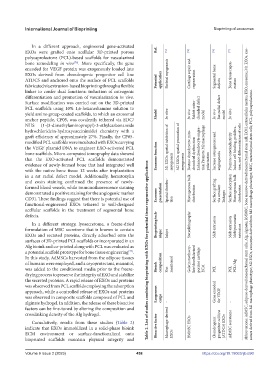
EXOs were grafted onto acellular 3D-printed porous Ref. [74] [75] [76] [77]
polycaprolactone (PCL)-based scaffolds for vascularized
bone remodeling in vivo . More specifically, the gene
[76]
encoded for VEGF protein was exogenously loaded into
EXOs derived from chondrogenic progenitor cell line Skeletal myogenesis Cartilage repair and Segmental bone Bone tissue regen-
ATDC5 and anchored onto the surface of PCL scaffolds Potential application regeneration
fabricated via extrusion-based bioprinting through a flexible defects eration
linker to confer dual functions: induction of osteogenic
differentiation and promotion of vascularization in vivo.
Surface modification was carried out on the 3D-printed
PCL scaffolds using 10% 1,6-hexanediamine solution to Rabbit osteo- chondral defect Rat radial defect
yield amino group-coated scaffolds, to which an exosomal Model In vitro In vivo model In vivo model In vitro
anchor peptide, CP05, was covalently tethered via EDC/
NHS (1-(3-dimethylaminopropyl)-3-ethylcarbonamide
hydrochloride/n-hydroxysuccinimide) chemistry with a
graft efficiency of approximately 27%. Finally, the CP05-
modified PCL scaffolds were incubated with EXOs carrying
the VEGF plasmid DNA to engineer EXO-activated PCL M1 EXOs: spatial inhibition of M2 EXOs: spatial promotion of Restore chondrocyte mito- Enhance chondrocyte migra- tion Promote M2 macrophage Induce cell binding, prolifera- tion, and differentiation of cells
bone scaffolds. Micro-computed tomography data showed Exosome function chondrial dysfunction Increase osteogenesis and Promote osteoinductivity
that the EXO-activated PCL scaffolds demonstrated myogenesis myogenesis polarization angiogenesis
evidence of newly-formed bone that had integrated well
with the native bone tissue 12 weeks after implantation
in a rat radial defect model. Additionally, hematoxylin
and eosin staining confirmed the presence of newly-
formed blood vessels, while immunofluorescence staining Homogenous bulk Surface adsorption Homogenous bulk
demonstrated a positive staining for the angiogenic marker Exosome presentation Spatial distribu- distribution Surface grafting via covalent distribution Abbreviations: AdMSC, adipose-derived mesenchymal stem cells; Alg, alginate; BMMSC, bone marrow-derived mesenchymal stem cell; ECM, extracellular matrix; EXO, exosome; M1 EXOs, exo-
CD31. These findings suggest that there is potential use of tion linkages
functional-engineered EXOs tethered to well-designed
acellular scaffolds in the treatment of segmental bone
defects. somes derived from M1 macrophage phenotype; M2 EXOs, exosomes derived from M2 macrophage phenotype; MSC, mesenchymal stem cells; PCL, polycaprolactone.
In a different strategy, lyosecretome, a freeze-dried Bioprinting tech- Stereolithography Melt/pneumatic
formulation of MSC secretome that is known to contain Melt extrusion Melt extrusion extrusion
EXOs and secreted proteins, directly adsorbed onto the nique Inkjet
surfaces of 3D-printed PCL scaffolds or incorporated in an
Alg bioink and co-printed along with PCL was evaluated as
a potential scaffold prototype for bone tissue engineering . Table 2. List of studies combining bioprinting with EXOs for potential tissue regenerative applications
[77]
In this study, AdMSCs harvested from the adipose tissues Gelatin methacry- late/decellularized porcine cartilage
of humans were employed, and a cryoprotectant, mannitol, composition mentioned
was added to the conditioned media prior to the freeze- Bioink Not ECM PCL PCL PCL/Alg
drying process to preserve the integrity of EXO and stabilize
the secreted proteins. A rapid release of EXOs and proteins
was observed from PCL scaffolds employing the adsorption
approach, while a controlled release of EXOs and proteins Exogenous Gene encoded
was observed in composite scaffolds composed of PCL and cargo for VEGF
alginate hydrogel. In addition, the release of these bioactive - - -
factors can be fine-tuned by altering the composition and
crosslinking density of the Alg hydrogel.
Cumulatively, results from these studies (Table 2) Macrophage-derived progenitor cell line AdMSC secretome
indicate that EXOs immobilized in a solid-phase bioink Bioactive factor BMMSC EXOs Chondrogenic (ATDC5) EXOs
ECM environment or surface-functionalized onto EXOs
bioprinted scaffolds maintain physical integrity and
Volume 9 Issue 2 (2023) 458 https://doi.org/10.18063/ijb.690