
Page 11 - IJB-9-3
P. 11
International Journal of Bioprinting Bioprinting of PDAC microtissues for drug screening
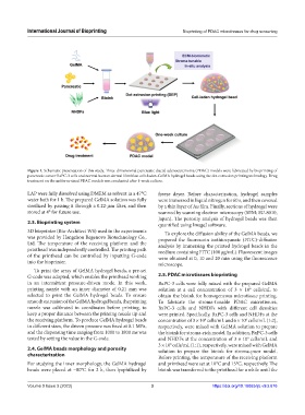
Figure 1. Schematic presentation of this study. Three-dimensional pancreatic ductal adenocarcinoma (PDAC) models were fabricated by bioprinting of
pancreatic cancer BxPC-3 cells and normal human dermal fibroblast cells laden-GelMA hydrogel beads using the dot extrusion printing technology. Drug
treatment on the uniform-sized PDAC models was conducted after 1-week culture.
LAP were fully dissolved using DMEM as solvent in a 47°C freeze dryer. Before characterization, hydrogel samples
water bath for 1 h. The prepared GelMA solution was fully were immersed in liquid nitrogen for 60 s, and then covered
sterilized by passing it through a 0.22-μm filter, and then by a thin layer of Au film. Finally, sections of hydrogel were
stored at 4° for future use. scanned by scanning electron microscopy (SEM; SU-8010,
Japan). The porosity analysis of hydrogel beads was then
2.3. Bioprinting system quantified using ImageJ software.
3D bioprinter (Bio-Architect WS) used in the experiments To explore the diffusion ability of the GelMA beads, we
was provided by Hangzhou Regenovo Biotechnology Co., prepared the fluorescein isothiocyanate (FITC)-diffusion
Ltd. The temperature of the receiving platform and the analysis by immersing the printed hydrogel beads in the
printhead was independently controlled. The printing path medium containing FITC (100 μg/mL). Fluorescent images
of the printhead can be controlled by inputting G-code were obtained at 0, 10 and 20 min using the fluorescence
into the bioprinter. microscope.
To print the array of GelMA hydrogel beads, a pre-set
G-code was adopted, which enables the printhead working 2.5. PDAC microtissues bioprinting
in an intermittent pressure-driven mode. In this work, BxPC-3 cells were fully mixed with the prepared GelMA
printing nozzle with an inner diameter of 0.21 mm was solution at a cell concentration of 3 × 10 cells/mL to
6
selected to print the GelMA hydrogel beads. To ensure obtain the bioink for homogeneous microtissue printing.
smooth extrusion of the GelMA hydrogel beads, the printing To fabricate the stroma-tunable PDAC microtissues,
nozzle was calibrated in coordinates before printing, to BxPC-3 cells and NHDFs with different cell densities
keep a proper distance between the printing nozzle tip and were printed. Specifically, BxPC-3 cells and NHDFs at the
the receiving platform. To produce GelMA hydrogel beads concentration of 3 × 10 cells/mL and 6 × 10 cells/mL (1:2),
6
6
in different sizes, the driven pressure was fixed at 0.1 MPa, respectively, were mixed with GelMA solution to prepare
and the dispensing time ranging from 1000 to 1800 ms was the bioink for stroma-rich model. In addition, BxPC-3 cells
tested by setting the value in the G-code. and NHDFs at the concentration of 3 × 10 cells/mL and
6
3 × 10 cells/mL (1:1), respectively, were mixed with GelMA
6
2.4. GelMA beads morphology and porosity solution to prepare the bioink for stroma-poor model.
characterization Before printing, the temperature of the receiving platform
For studying the inner morphology, the GelMA hydrogel and printhead were set at 10°C and 15°C, respectively. The
beads were placed at −80°C for 2 h, then lyophilized by bioink was transferred to the printhead for a while until the
Volume 9 Issue 3 (2023) 3 https://doi.org/10.18063/ijb.v9i3.676