
Page 323 - IJB-9-3
P. 323
International Journal of Bioprinting New fibrillar collagen for 3D printing and bioprinting
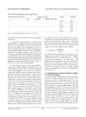
Table 1. Bioink concentration, cell type, and bioink code
Final ColN concentration (% w/w) Mixture ratios (mL) Cell type Bioink code
ColN TRIS-HCl DMEM (w and w/ cells)
2.0 2 1.5 1.5 MSC-D1 2MSC
L929 2L929
No cells 2CTR
3.0 3 1 1 MSC-D1 3MSC
L929 3L929
No cells 3CTR
In all cases, the cellular density was maintained at 2 × 10 cell/mL.
5
steps indicated in the ISO 10993-5 for direct and indirect in a microplate reader (Epoch, BioTek). The results are
cytotoxicity. expressed as percentage of cell viability with respect to the
positive control (Equation I). OD refers to the average
A collagen film of approximately 2 mm thickness was 440s
prepared from ColA, followed by NaOH neutralization (50 O.D. value of the test sample, while OD 440b stands for the
blank, empty well. The cytotoxicity of the sample is inversely
mM, 30 min). The resultant, neutral film was rinsed with
PBS twice (15 minutes each) to eliminate any excess of proportional to the percentage of cellular viability.
NaOH. Afterward, the neutral ColA film was submerged in 100 × OD
complete DMEM supplemented with 1% (v/v) of penicillin/ Cellviability % = OD 440 s (I)
streptomycin overnight. Rounded scaffolds with 1 cm 440 b
2
surface were cut from the neutral collagen film and deposited The viability of L929 cells seeded on the surface of each
in a 48-well plate. With respect to the neutral formulation, collagen mass was confirmed with a Live/Dead Viability/
TM
ColN collagen ink was neutralized by following the same Cytotoxicity Kit, based on calcein-AM (green, live cells)
procedure explained elsewhere (TRIS-HCl, 1.5 M, pH 7.4). and ethidium homodimer-1 (red, dead cells). Cell-
Afterwards, corresponding DMEM amount was mixed up seeded scaffolds were treated according to manufacturer’s
to 40 times to ensure a correct homogenization. instructions, and samples were imaged with an inverted
fluorescence microscope (Nikon, AZ100). The split channel
In order to quantify cellular viability, cell proliferation
reagent WST-1 (Merck, Germany) was employed. WST-1 is images were subsequently merged by ImageJ® software
a water-soluble tetrazolium salt (pink) that is cytosolically (Fiji). This experiment was carried out in duplicates for
reduced by dehydrogenases into a formazan dye (yellow/ each time point.
orange) with absorbance peak at 440 nm, whose value is 2.6. 3D bioprinting and culture of cell-laden scaffolds
directly proportional to the amount of living cells. For 2.6.1. Bioink formulation
the direct assay, L929 fibroblasts were incubated with a Six different collagen bioinks were prepared starting from
4
2
density of 3.12 × 10 cells/cm for 24 h until fully confluent. ColN in an attempt to evaluate the performance of this
Subsequently, acid and neutral gelified collagen inks at formulation during 3D bioprinting (Table 1). The acid
2% (w/w) and 3% (w/w) were introduced within the cell- mass (5% w/w), sterile ColN at room temperature was
seeded wells for 24 h, which were later removed, and the mixed with TRIS-HCl buffer (1.5 M, pH 7.5–7.6, sterilized
WST-1 solution (1:44 dilution) was added for 1 h at cell by filtration with 0.2-μm nitrocellulose filters) by passing
culture conditions. Analogously, for the indirect assay, non- both ingredients (see Table 1 for ratios) from one syringe
laden collagen samples were introduced in empty wells to another (Luer slip and Luer lock syringes, B Braun ),
TM
and covered in DMEM for 24 h. The resulting exudate connected though a Luer lock connector. A total of 40 passes
was transferred to individual wells seeded with L929 cells is enough to guarantee total collagen neutralization and
(3.12 × 10 cells/cm ) and incubated for 24 h. Then, the homogeneous mixture. Right after this, the corresponding
2
4
medium was removed, and the WST-1 cytotoxicity assay amount of culture medium (see Table 1 for ratios, with
was performed as indicated above. All samples were done or without MSC-D1 or L929 cells suspended) is mixed
in triplicates with Latex® 1 cm square membranes used with the neutralized ColN (approximately 20–25 syringe
2
as negative, cytotoxic controls, whereas positive controls passes). Both the buffer and the collagen neutralization
consist of L929 fibroblasts with standard DMEM. Optical process are performed extemporaneously right before 3D
density (O.D.) of WST-1 reagent was measured at 440 nm
Volume 9 Issue 3 (2023) 315 https://doi.org/10.18063/ijb.712