
Page 327 - IJB-9-3
P. 327
International Journal of Bioprinting New fibrillar collagen for 3D printing and bioprinting
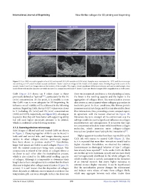
Figure 3. Cryo-SEM microphotographs of acid (A) and neutral (B) ColN samples at 0.5% (w/w). Samples were maintained at -50°C and the microscope
was working at 5 kV under medium vacuum conditions. Insets 1 and 4 are images of the middle of the sample mass, showing the general texture of the
sample; insets 2 and 5 are images taken at the borders of the samples. This region reveals significant differences between acid and neutral sample, since
looser fibers with smaller diameter are visible in inset 2 in comparison with inset 5. Insets 3 and 6 are detailed images (800× magnification in both cases).
ColN (Figure 2D) shows tan δ values closer to those these environmental conditions to the physiological ones,
[32]
previously defined as “optimal” , particularly for the 2% the lower is the swelling capacity and the higher is the
(w/w) concentration. At this point, it is possible to state aggregation of collagen fibers. The neutralization process
that ColN mass is more adequate for 3D bioprinting. Its also creates an environment where collagen approaches its
influence on cell viability will be addressed in the following isoelectric point. In these conditions, this fibrous protein
sections. Regarding ColA, the tan δ (G*) values were closer possesses neutral net charge, and this may also enable closer
to 0 (including 4% [w/w] and 5% [w/w] concentrations fiber interactions, thus promoting a more compact aspect,
[0.082 and 0.078, respectively, see Figure S2]), allowing us in agreement with the texture observed in Figure 3B.
to predict that they will have better self-supporting ability Moreover, the ionic strength of the environment and the
but will need higher pneumatic pressure to be printed, salt type could also exert a significant influence on collagen
which is confirmed in the following sections. microstructure and entanglement. It is known that high
ion concentration leads to water withdrawal from collagen
3.1.2. Scanning electron microscopy molecules, which promotes more collagen–collagen
SEM images of diluted acid and neutral ColN are shown interactions (predominant hydrophobic interactions) .
[34]
in Figure 3. Clumping together of fibrils can be found in
both acid and neutral inks, and images showing aspect Higher apparent viscosities have been reported for acidic
similar to other collagen electron microscopy images ColA ink with respect to neutral ColN (Figure 2). That
have been shown in previous studies [25,33] . Nonetheless, is, it is expected that more fiber interactions would lead to
larger void spaces are visible in acid collagen (Figure 3A), higher viscosities. Nonetheless, we observed the contrary.
with the neutral counterpart being more compact. This Inconsistences in rheological behavior of type I collagen
[27]
compactness is related to the ability of collagen fibers to have already been reported . In the acidic ColA ink, fibrils
be closer to each other after the neutralization process, are swollen up to its maximum capacity and are more freely
which is also related to the differential swelling capacity and homogeneously distributed in the whole collagen ink,
of collagen. Although it is impossible to determine their which enables them to actively participate in the formation
length due to their entanglement, it is evident that the fiber’s of an internal network that exerts higher resistance to
diameter is higher after collagen neutralization (Figure 3B, external stresses (higher viscosity). It can be hypothesized
arrows). It is well known that the disposition of collagen that the neutralization process changes the environment
fibers depends on different environmental conditions like and induces some release of water from collagen fibrils,
temperature, pH, and ionic strength. In fact, the closer are which may aggregate between each other. Under these
Volume 9 Issue 3 (2023) 319 https://doi.org/10.18063/ijb.712