
Page 329 - IJB-9-3
P. 329
International Journal of Bioprinting New fibrillar collagen for 3D printing and bioprinting
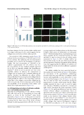
Figure 5. Cell viability (%) of L929 fibroblast seeded on ColA (A1 and A2) and ColN (B1 and B2) inks according to WST-1 (A, B) and Live/Dead assays
(1–2). Scale bars: 200 µm.
have been detected, the fact that the cellular viability tend printing nozzle’s wall, the flow is faster and the shear stress
to be higher with respect to the control suggests that the is higher, which causes cell deformation and subsequent
collagen inks could induce or favor cellular growth. death . Notwithstanding this fact, the bioprinting process
[35]
herein reported can be considered harmless for L929
Particularly for ColN, cytotoxicity results (Figure 5B)
indicate that the concentration of TRIS-HCl used (1.5 M) and MSC-D1 since (as shown by the cell viability results
represented in Figure 8), 90% of cellular viability has
is non-cytotoxic, thus indicating that the neutralization been reported at day 0 in all cases. Moreover, living cells
procedure can be used for the formulation of cell-laden are homogenously distributed throughout all the printing
bioinks. The final concentration of TRIS-HCl in the lines, thus suggesting that the diffusion of nutrients and
scaffold is approximately 250 mM, which is less than oxygen inside the bioink network is sufficient to guarantee
the concentration of other medical materials approved cellular growth.
by the FDA and clinically available, like Tromethamine
injection which is used to prevent and treat severe The cellular viability has been quantified based on
[37]
metabolic acidosis. In addition, the concentration of this analysis with results shown in Figure 8. Nevertheless,
collagen does not appear to be a parameter affecting cell the unreliability of the quantification methodology
viability. Therefore, any differences on cellular viability increases with time, since cells start to confluence and to
detected after the 3D bioprinting of neutral collagen bioinks become indistinguishable, as easily observable in some
would probably be related to the bioprinting conditions of the Live/Dead images at day 12 (Figure 7A, day 12).
rather than to the collagen concentration. Regarding the This fact must be taken into consideration during the
ColA ink (acidic ink, 5% w/w), these tests confirm the full interpretation of results, which means that the cellular
biocompatibility of the formulation (both indirect and viability reported for days 6 and 12 could be higher
direct) after simple neutralization (Figure 5A). than estimated. Fibroblasts maintained a constant and
high viability throughout the whole experiment (≥90%)
3.2. 3D bioprinting and culture of cell-laden scaffolds (Figure 8A). No significant differences have been found
3.2.1. Cell viability and proliferation between different collagen concentrations. This is in
Right after the 3D bioprinting of ColN, cell-laden bioinks, agreement with the direct and indirect cytotoxicity tests
dead cells (red) are by far outnumbered by living cells mentioned in section 3.1.4 (Figure 5) and even with some
(green), as shown by the Live/Dead assay results results in the literature from collagen bioinks within
TM
presented in Figures 6 and 7. At this point, the majority the same order of concentration [24,38] . It is also worth to
of dead cells are located on the verge of the printing lines, mention that the collagen used by Osidak et al. was a
[28]
revealing that the shear stress of the bioink extruded soluble collagen that required fibrillogenesis triggered
through the printing gauge induces cell death. Next to the
by TRIS-HCl neutralization at 4°C and then collagen
Volume 9 Issue 3 (2023) 321 https://doi.org/10.18063/ijb.712