
Page 332 - IJB-9-3
P. 332
International Journal of Bioprinting New fibrillar collagen for 3D printing and bioprinting
Figure 8. Cell viability (%) of fibroblasts (A) and mesenchymal cells (B) cultured within the bioprinted scaffolds. Results are based on Live/Dead assay.
TM
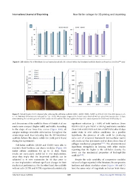
Figure 9. Optical density (O.D.) obtained after culturing the cell-laden scaffolds (2MSC, 2L929, 3MSC, 3L929) in 10% of CCK-8 for 24 h (mean ± s.d.;
n = 6). Statistical differences are indicated by * (p < 0.05). Microscopic images at the bottom were obtained with an optical microscope after 12 days,
demonstrating the extensive growth of L929 outside the 3D scaffold. This fact explains the high O.D. values reported for 2L929 and 3L929 at day 12.
and dimensions of the scaffolds: those of Osidak et al. are significant reduction (p < 0.05) of both hardness (from
much more compact (higher infill) and width. According 922.03 ± 22.15 g to 762.67 ± 105.9 g) and elastic modulus
to the shape of our force-time curves (Figure 10A), all (from 0.48 ± 0.0219 kPa to 0.364 ± 0.0479 kPa) after 12 days
samples undergo reversible deformation throughout the under static in vitro culture conditions. As a possible
strain range used, thus indicating that the 3D-bioprinted hypothesis, the presence of cells could be producing
scaffolds behave like elastic solids (no yield point found some sort of structural framework (extracellular matrix
during the experiment). sub-products) that minimizes and/or slows down loss of
Cell-laden scaffolds (2L929 and 3L929) were able to collagen mechanical properties [39-43] . The aforementioned
maintain their hardness and elastic modulus (Figure 10) hypothesis strengthens in keeping with other studies
under culture conditions for up to 12 days. These reporting that the higher is the cell-laden density, the
results are particularly useful for in vivo implantation, lower are the mechanical properties of hydrogel-like
[44-46]
since they imply that the bioprinted scaffolds can be scaffolds .
subjected to in vitro maturation for 12 days prior to Despite the wide variability of compressive modulus
in vivo implantation without significant changes in their values of collagen reported in the literature, the compressive
mechanical performance. On the other hand, the scaffolds hardness and elastic modulus values (Figure 10B and C)
without cells (2CTR and 3CTR) experienced a statistically have the same order of magnitude as human brain tissue
Volume 9 Issue 3 (2023) 324 https://doi.org/10.18063/ijb.712