
Page 330 - IJB-9-3
P. 330
International Journal of Bioprinting New fibrillar collagen for 3D printing and bioprinting
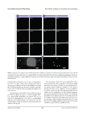
Figure 6. Fluorescence microscopy for the (A) 2MSC and (B) 3MSC scaffolds. Live cells (green) and dead cells (red) were stained with calcein-AM and
ethidium homodimer-1, respectively. These microphotographs were obtained by performing z-stack of the scaffolds and merging both channels (4×
magnification). (A and B) The scale bar corresponds to 200 µm. (C) Detailed image at day 0 of the 3D-printed scaffold merging brightfield, calcein-AM,
and ethidium homodimer-1 channels. Yellow arrows (left) point to air bubbles in the bioink; white arrows (right) mark the location of dead cells in the
printing line edge.
polymerization induced by the increase in temperature The absorbance values (O.D.) generated by the CCK-8
(at 37°C). In other words, these authors were able to tests are rather low when compared to the results usually
extrude type I collagen with good cell viability results, but obtained in 2D cultures. This fact can be explained by the
5
the 3D bioprinting process was more complex, needing a low cellular density within the bioinks (2 × 10 cell/mL)
careful control of factors like temperature throughout the and the 3D nature of the scaffolds. In these conditions,
entire process. the WST-8 must reach the cells embedded within the 3D
scaffold and be metabolized, and the resultant formazan
The metabolic activity of MSC-D1 and L929 determined
by CCK-8 is shown in Figure 9. CCK-8 contains WST- dye must be released to the culture medium to be effectively
quantified. In 2D cultures, the CCK-8 test takes about 4 h
8 (a water-soluble tetrazolium salt, yellow) that can be to complete. Nonetheless, it has been observed that 4 h are
reduced by dehydrogenase in the cell to a corresponding not enough to achieve reliable and reproducible O.D. data
formazan dye (yellow/orange) that absorbs at 450 nm. from the 3D collagen scaffolds. Instead, the CCK-8 assay
The absorbance of the formazan salt is proportional to the was left in contact with the scaffolds for 24 h.
number of living cells.
Volume 9 Issue 3 (2023) 322 https://doi.org/10.18063/ijb.712