
Page 342 - IJB-9-3
P. 342
International Journal of Bioprinting Amniotic fornical ring for ocular surface reconstruction
SJS and TEN with extended ocular surface damage. Here, (P = 0.812, Table 2). In our study, the AM dissolution
we provided some eye pictures of patients with AFR, time was shorter than that reported in the literature .
[5]
including TEN, thermal burn, acid burn, and alkaline The thickness of the AM was also one of the main
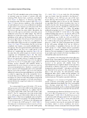
burns (Figure 2). Moreover, we showed a long follow- factors affecting AM dissolution . AFR and suture had
[2]
up of patients with TEN who received AFR (Figure 2C). similar dissolution times as that for the AM. Therefore,
Figure 2A shows extensive exudation in the conjunctival we speculated that the shorter dissolution time may be
sac and widespread ocular surface epithelial and palpebral related to the thin AM. The percentages of healed surface
margin defects in an intensive care unit patient with TEN area were 90.91% (66.10%–100.00%) for AFR and 93.67%
before AFR-assisted AMT. AFR supported the AM to (60.23%–100.00%) for SAMT (P = 0.994). The median
the fornices and kept the eyelid closed. Meanwhile, the epithelial healing time was 14 (7–75) days for AFR and 30
AM covered the superior and inferior eyelid skin several (14–55) days for SAMT (P = 0.436, Table 2). The average
millimeters away from the eyelid margin. The AM had epithelial healing time was 44 ± 52 days for AFR and 40
dissolved at 3 weeks after surgery in two eyes. The corneal ± 41 days for SAMT (P = 0.751, Table 2). The incidence
epithelium of the right eye had healed completely, while of symblepharon was 31.58% for AFR and 40.91% for
the corneal epithelium of the left eye still had a flaky defect SAMT (P = 0.465). Limbal stem cell deficiency accounted
at 3 weeks after surgery. At this time, the epithelium of for 31.58% in the AFR group and 54.55% in the SAMT
the double eyelid margin was still not completely repaired group (P = 0.080). No conjunctival granulomas occurred
(Figure 2A). Figure 2B demonstrates that the cornea was in the AFR group, although there was no difference
completely clear despite a punctured epithelial defect in in the incidence of granulomas between the AFR and
the right eye. The cornea presented with scattered punctate SAMT groups (P = 0.407). Suture-induced conjunctival
epithelial defects, and there was a slight opacity in the granuloma is a chronic inflammation caused by suture
left eye at 3 months after the operation (Figure 2B). At irritation. The lower incidence of conjunctival granulomas
the same time, the eyelid margin also showed moderate in our study may be related to the faster dissolution of the
congestion and inflammation (Figure 2B). The cornea AM and the earlier removal of sutures.
of the right eye was clear, and the corneal opacity of the
left eye was significantly reduced 9 months after surgery Two patients in the AFR group suffered from glaucoma
(Figure 2C). The best corrected VA was 1.0 for the right eye complications. Three patients in both the AFR and SAMT
and 0.5 for the left eye 9 months after surgery. However, groups developed traumatic cataracts. These complications
ProKera, another sutureless AM mounted device, is occurred in the patients with alkaline burns. We
approximately 16 mm in diameter and can only cover the hypothesized that this might be related to the high tissue
cornea and peripheral sclera. Therefore, it cannot be used permeability of alkaline fluid and the administration of
[17]
alone in patients with widespread ocular surface and eyelid postoperative glucocorticoids . Subconjunctival effusion
injuries, such as SJS and TEN . AFR can be used alone for was observed in two patients in the acute stage of burn
[5]
widespread ocular surface and eyelid injuries, and its effect injury in the AFR group. The subconjunctival effusion
is related to supporting AM to the conjunctival fornix. disappeared after correct ocular nursing guidance. All
It may be that just the AFR had the ability to extend and enrolled patients tolerated the device during the whole
immobilize the AM more fully. Three patients complained treatment process. We found no associated infections.
of mild foreign body sensation in the early postoperative Subsequent to the removal of 3D-printed AFR, one
period, followed by a significant reduction in discomfort patient underwent penetrating keratoplasty, and eight
and disappearance. patients underwent tarsorrhaphy. The other nine patients
wore a bandage contact lens (Table 1). Three patients
The median initial VA was 0.959 (0.600–1.775) for the underwent tarsorrhaphy, and four patients needed bandage
AFR group and 1.150 (0.700–2.000) for the SAMT group. contact lens treatment after the removal of the sutured AM.
During post-treatment follow-up, the median final VA
was 0.300 (0.100–0.900) (AFR) and 0.450 (0.300–1.200) The mean operative duration (MOD) per eye was 18.4 ±
(SAMT). The VA improvement was 0.400 (0.200–0.900) 10.1 min in the AFR group and 42.2 ± 18.5 min in the
in the AFR group and 0.500 (0.200–0.800) in the SAMT SAMT group (P = 0.000, Table 3). The MOD per eye was 16
group (Table 2). There was no significant initial or final VA (10–24) min in the AFR group and 35 (30–47) min in the
difference between the two groups. The median dissolution SAMT group (P = 0.000, Table 3). There were significant
or removal times were 7 (7–19) days in the AFR group and differences in the MOD between the two groups.
14 (7–14) days in the SAMT group (P = 0.812, Table 2). Shorter operative times may be associated with bedside
The mean time of dissolution or removal was 15 ± 11 days procedures, topical or surface anesthesia, and suture-
in the AFR group and 14 ± 7 days in the SAMT group free techniques. Several techniques have been previously
Volume 9 Issue 3 (2023) 334 https://doi.org/10.18063/ijb.713