
Page 340 - IJB-9-3
P. 340
International Journal of Bioprinting Amniotic fornical ring for ocular surface reconstruction
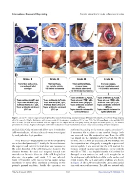
Figure 1. (A) The 3D-printed design and a photograph of the amniotic fornical ring. The physical image of trimmed 3D-printed AFR; software design diagram
of AFR; image of AFR after disinfection with ethylene oxide. (B) Implantation procedures of 3D-printed AFR. The AFR was placed on the unfolded AM
(40 × 60 mm). The AM with an enclosed AFR was slipped into the conjunctival sac after gently turning the upper and lower eyelids. (C) The removal
procedures of AFR. (D) Diagram outlining the protocol for medication of ocular manifestations in acute ocular burns according to Roper-Hall grades.
and July 2022. Only patients with follow-up ≥ 2 weeks after performed according to the routine aseptic procedure .
[11]
AM were included. Written informed consent was signed If necessary, the exudate or any residual foreign body
by each patient or legal guardian. was removed from the conjunctival sac. Then, the AFR
was placed on the expanded cryopreserved AM (40 ×
First, the doctor measured the size of the conjunctival 60 mm). The AM with an enclosed AFR was inserted into
sac as described previously . Briefly, the distance between the conjunctival sac after gently turning the superior and
[10]
the superior and inferior fornical rims was measured as inferior eyelids. It was ensured that the AFR reached the
the outer diameter of the AFR transverse diameter. The fornices without causing lagophthalmos. In the patients
distance between the nasal and temporal fornical rims with SJS or TEN, it was required that the AM covered the
was taken as the outer diameter of the AFR longitudinal damaged cornea, conjunctiva, and eyelid margin due to
diameter. Appropriate and sterile AFR was selected. the widespread epithelial defects of the ocular surface and
Next, AFR-assisted AMT was performed under surface eyelid margin. The AFR application methods are shown
anesthesia or nerve block anesthesia depending on the in Figure 1B. The AM was anchored to the external eyelid
patient’s general condition. Briefly, the operation was skin with sutures if necessary. The AM was sutured with
Volume 9 Issue 3 (2023) 332 https://doi.org/10.18063/ijb.713