
Page 343 - IJB-9-3
P. 343
International Journal of Bioprinting Amniotic fornical ring for ocular surface reconstruction
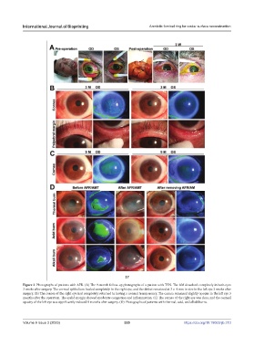
Figure 2. Photographs of patients with AFR. (A) The 9-month follow-up photographs of a patient with TEN. The AM dissolved completely in both eyes
3 weeks after surgery. The corneal epithelium healed completely in the right eye, and the defect remained at 3 × 4 mm in size in the left eye 3 weeks after
surgery. (B) The cornea of the right eye had completely returned to having a normal transparency. The cornea remained slightly opaque in the left eye 3
months after the operation. The eyelid margin showed moderate congestion and inflammation. (C) The cornea of the right eye was clear, and the corneal
opacity of the left eye was significantly reduced 9 months after surgery. (D) Photographs of patients with thermal, acid, and alkali burns.
Volume 9 Issue 3 (2023) 335 https://doi.org/10.18063/ijb.713