
Page 178 - IJB-9-4
P. 178
International Journal of Bioprinting Multi-scale vascularization strategy for 3D-bioprinted tissue
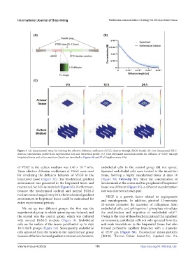
Figure 3. (A) Experimental setup for deriving the effective diffusion coefficient of FITC-dextran through dECM bioink. (B) One-dimensional FITC-
dextran concentration profile from experimental data and theoretical profile. (C) Time-dependent simulation results for diffusion of VEGF through
bioprinted tissue and culture medium (details are described in Figures S6 and S7 of Supplementary File).
of VEGF in the culture medium was 1.46 × 10 m /s. endothelial cells in the control group did not sprout.
−10
2
These effective diffusion coefficients of VEGF were used Sprouted endothelial cells were located in the interstitial
for simulating the diffusion behavior of VEGF in the tissue, forming a highly vascularized tissue at days 14
bioprinted tissue (Figure 3C). The biochemical gradient (Figure 5B; Videoclip S1). Since the concentration of
environment was generated in the bioprinted tissue and biochemicals at the center and the peripheral of bioprinted
maintained for 24 h as intended (Figure S5). Furthermore, tissue was different (Figure S5), a different vascularization
because the biochemical cocktail and normal EGM-2 rate was observed on each part.
medium were changed every 24 h, the biochemical gradient VEGF is a growth factor related to angiogenesis
environment in bioprinted tissue could be maintained for and vasculogenesis. In addition, phorbol 12-myristate
entire experimental periods.
13-acetate promotes the secretion of collagenase from
We set up two different groups: the first was the endothelial cells, and sphingosine-1-phosphate stimulates
[7]
experimental group in which sprouting was induced, and the proliferation and migration of endothelial cells .
the second was the control group, which was cultured Owing to the roles of these biochemicals and their gradient
with normal EGM-2 medium (Figure 4). Endothelial environment, endothelial cells not only sprouted from the
cells on the surface of the lumen proliferated up to days mid-scale vasculatures in the bioprinted tissue, but also
10 in both groups (Figure 5A). Subsequently, endothelial formed perfusable capillary branches with a diameter
cells sprouted from the lumen in the experimental group of 19.57 μm (Figure S6). Fluorescence micro-particles
because of the biochemical gradient environment; however, (R0100, Thermo Fisher Scientific), injected into the
Volume 9 Issue 4 (2023) 170 https://doi.org/10.18063/ijb.726