
Page 179 - IJB-9-4
P. 179
International Journal of Bioprinting Multi-scale vascularization strategy for 3D-bioprinted tissue
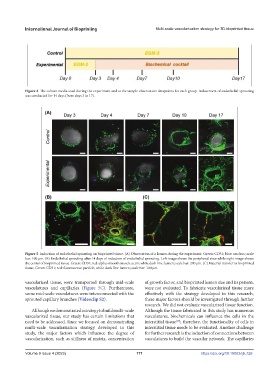
Figure 4. The culture media used during the experiment and at the sample observation timepoints for each group. Inducement of endothelial sprouting
was conducted for 14 days (from days 3 to 17).
Figure 5. Induction of endothelial sprouting on bioprinted tissue. (A) Observation of a lumen during the experiment. Green: CD31; blue: nucleus; scale
bar: 100 μm. (B) Endothelial sprouting after 14 days of induction of endothelial sprouting. Left image shows the peripheral view while right image shows
the center of bioprinted tissue. Green: CD31; red: alpha-smooth muscle actin; white dash line: lumen; scale bar: 200 μm. (C) Material transfer in bioprinted
tissue. Green: CD31; red: fluorescence particle; white dash line: lumen; scale bar: 100 μm.
vascularized tissue, were transported through mid-scale of growth factor, and bioprinted lumen size and its pattern,
vasculatures and capillaries (Figure 5C). Furthermore, were not evaluated. To fabricate vascularized tissue more
some mid-scale vasculatures were interconnected with the effectively with the strategy developed in this research,
sprouted capillary branches (Videoclip S2). these major factors should be investigated through further
research. We did not evaluate vascularized tissue function.
Although we demonstrated a strategy to build multi-scale Although the tissue fabricated in this study has numerous
vascularized tissue, our study has certain limitations that vasculatures, biochemicals can influence the cells in the
need to be addressed. Since we focused on demonstrating interstitial tissue ; therefore, the functionality of cells in
[19]
multi-scale vascularization strategy developed in this interstitial tissue needs to be evaluated. Another challenge
study, the major factors which influence the degree of for further research is the induction of connections between
vascularization, such as stiffness of matrix, concentration vasculatures to build the vascular network. The capillaries
Volume 9 Issue 4 (2023) 171 https://doi.org/10.18063/ijb.726