
Page 103 - IJB-9-5
P. 103
International Journal of Bioprinting Application and prospects of 3D printable microgels
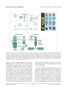
Figure 4. 3D Bioprinting of macroporous materials based on entangled hydrogel microstrands. (A) Sizing of the bulk tyramine-modified hyaluronic
acid (HA-TYR) hydrogels using grids with aperture diameter of 40, 100, and 500 µm. (B, C) 3D printing schematic of HA-TYR bioinks. The cell-laden
microgels can be printed through a 0.61-mm nozzle and stabilized by secondary crosslinking [(from ref. licensed under Creative Commons Attribution
[96]
4.0 license)]. (D) The bulk hydrogel forms a microchain structure after mechanical crushing, and has moldability, stability in aqueous solutions, porosity,
printability. (E) A bioink can be created by embedding cells in bulk hydrogel before sizing that results in a spatial deposition of cells inside the gel phase.
Alternatively, cells can be mixed in between the already prepared entangled microstrands, so cells occupy the space outside the gel phase [(from ref.
[97]
licensed under Creative Commons Attribution 4.0 license)].
networks: a solid but brittle microgel network and a soft that the mechanical properties of microgels are sufficient to
hydrogel network. As a result, this P-DN microgel exhibits meet the needs of most 3D bioprinting, while also having
exceptional stretchability. The viscosity, yield stress, and strong customizability.
strength of this microgel increase with increasing particle
concentration, and its fracture toughness is capable of The flow of hydrogel–microgel during the process of
reaching 3000 J m −2[98] . Seymour et al. reported a strategy 3D bioprinting differs significantly from the liquid flow of
involving the mixing of two microgels (GelMA: gelatin) ordinary hydrogels. In order to achieve an ideal printing
and the removal of the gelatin microgel after crosslinking, effect, there are still many challenges to be addressed.
which resulted in a final microgel with controllable pore To address this issue, Xin et al. utilized thiol-epoxy PEG
size by manipulating the mixing ratio and diameter of the microgel as biological ink and studied ways to improve
two microgels . According to the experimental results, printing performance . They first constructed microgels in
[89]
[67]
these microgels showed a decrease in storage modulus as three different sizes (single disperse 100 μm, single disperse
the pore ratio increased, with the highest (pure GelMA 150 μm, and multiple disperse 200 μm) for testing and
microgel) and lowest ratio (40:60) storage moduli being found that single disperse microgel could be easily printed
4.5 ± 1.0 kPa and 177 ± 26 Pa, respectively . It can be seen by nozzles with diameters of 200–400 μm, while multiple
[89]
Volume 9 Issue 5 (2023) 95 https://doi.org/10.18063/ijb.753