
Page 433 - IJB-9-5
P. 433
International Journal of Bioprinting 3D printed topographically fabricated micron track peripheral nerve conduit
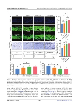
Figure 4. Histological characterization of peripheral nerve regeneration promoted by CC, MTC, MTC@NT3, and Autologous. (A) NF200
immunofluorescence staining to observe axonal regeneration. (B) The measured optical density of NF200 staining in the respective groups. (C)
Toluidine blue staining of semithin sections showing nerve regeneration. (D) The counted density of myelinated nerve fibers in the respective groups. (E)
Electrophysiological tests of regenerated nerves. (F, G) CAMP amplitude and CAMP latency statistics of regenerated nerves. (ns = not significant, *P <
0.05, **P < 0.01.)
group and the MTC@NT3 group had a higher density group and the CC group, while the MTC@NT3 group
of axons, suggesting that the regenerated nerve had a was able to promote the regeneration of axons more
better organization (Figure S4 in Supplementary File). In significantly (Figure 4B). In week 12, we saw a similar
addition, we also counted the visual density of regenerated pattern (Figure S6 in Supplementary File). Many studies
axons in immunofluorescence staining using ImageJ. The have realized that Schwann cells and axons react as a unit
results show no significant difference between the MTC and that changes in Schwann cells cause corresponding
Volume 9 Issue 5 (2023) 425 https://doi.org/10.18063/ijb.770