
Page 436 - IJB-9-5
P. 436
International Journal of Bioprinting 3D printed topographically fabricated micron track peripheral nerve conduit
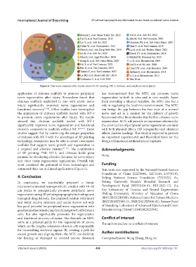
Figure 6. This work compares with studies related to 3D printing, NT-3, chitosan, and peripheral nerve injury.
application of chitosan scaffolds to promote peripheral has demonstrated that the MTC can promote nerve
nerve regeneration after injury. Researchers found that regeneration in both in vitro and in vivo models. Apart
chitosan scaffolds implanted in rats with sciatic nerve from providing a physical template, the MTC also has a
injury significantly improved nerve regeneration and role in regulating the local microenvironment. The MTC
functional recovery [73,74] . Other studies have investigated can bridge the gap between the two ends of a severed
the application of chitosan scaffolds loaded with NT-3 nerve and act as a conduit for the delivery of growth
to promote nerve regeneration after injury. The results factors and other biomolecules that further enhance nerve
showed that chitosan scaffolds loaded with NT-3 regeneration. MTC will provide an important reference for
significantly improved nerve regeneration and functional the construction of peripheral nerve regeneration conduits
recovery compared to scaffolds without NT-3 [75,76] . These with both physical effects (3D topography) and chemical
studies suggest that by combining the unique properties effects (factors-loading). This study is expected to provide
of chitosan with NT-3 with the advantages of 3D printing an important experimental and theoretical basis for the
technology, researchers may be able to create customized design of functional artificial neural implants.
scaffolds that support nerve growth and regeneration in
a targeted and effective manner [77–79] . The combination Acknowledgments
of 3D printing, PNI, NT-3, and chitosan holds great
promise for developing effective therapies for nerve injury None.
and other tissue regeneration applications. Overall, this
work combined the potential of these technologies and Funding
optimized their use in clinical applications (Figure 6). This work was supported by the National Natural Science
Foundation of China (22278003, 52273120, 21975019),
4. Conclusion Beijing National Science Foundation (7212121), the
In conclusion, we successfully prepared a bionic Peking University People’s Hospital Research and
microenvironmental neuroprosthetic conduit with 10–30 Development Fund (RDH2020-01, RDL2022-17), the
μm tracks to synergistically promote peripheral nerve Key Laboratory of Trauma and Neural Regeneration
regeneration using 3D printing topography technology and (Peking University), Ministry of Education of China
biological drug delivery. The prepared conduit with intact (BMU2022JDJS008), National Center for Trauma Medicine
and stable micron structure and neural factors not only (BMU2020XY005-01, BMU2021XY008-01), Science Fund
has good potential for peripheral nerve regeneration with of Shandong, Laboratory of Advanced Materials and Green
good properties of inducing directional growth of Schwann Manufacturing (Yantai) (AMGM2023F04).
cells, but also significantly promotes the regeneration
and functional recovery of axons. The channels on MTC Conflict of interest
serve as a physical guide for the regeneration of axons, The authors declare no conflicts of interest.
which are the lengthy extensions of nerve cells responsible
for transmitting electrical signals. By creating a path for Author contributions
axonal growth and aligning them, the MTC can facilitate
the healing of damaged or severed nerves. Research Conceptualization: Meng Zhang, Heng An
Volume 9 Issue 5 (2023) 428 https://doi.org/10.18063/ijb.770