
Page 488 - IJB-9-5
P. 488
International Journal of Bioprinting CECM-GelMA bioinks of DLP 3D printing for corneal engineering
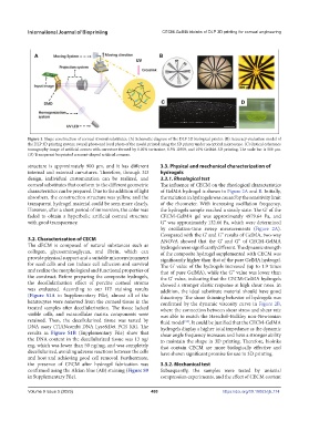
Figure 1. Shape construction of corneal stromal substitutes. (A) Schematic diagram of the DLP 3D biological printer. (B) Accuracy evaluation model of
the DLP 3D printing system; overall photo and local photo of the model printed using the 3D printer under an optical microscope. (C) Optical coherence
tomography image of artificial cornea with curvature formed by 0.02% tartrazine, 0.5% I2959, and 10% GelMA 3D printing. The scale bar is 500 μm.
(D) Transparent bioprinted crescent-shaped artificial corneas.
structure is approximately 900 μm, and it has different 3.3. Physical and mechanical characterization of
internal and external curvatures. Therefore, through 3D hydrogels
design, individual customization can be realized, and 3.3.1. Rheological test
corneal substitutes that conform to the different geometric The influence of CECM on the rheological characteristics
characteristics can be prepared. Due to the addition of light of GelMA hydrogel is shown in Figure 2A and B. Initially,
absorbers, the construction structure was yellow, and the the variation in hydrogels was caused by the sensitivity limit
transparent hydrogel material could be seen more clearly. of the rheometer. With increasing oscillation frequency,
However, after a short period of immersion, the color was the hydrogels sample reached a steady state. The G’ of the
faded to obtain a hyperbolic artificial corneal structure CECM-GelMA gel was approximately 4979.64 Pa, and
with good transparency. G” was approximately 132.66 Pa, which were determined
by oscillation-time sweep measurements (Figure 2A).
Compared with the G’ and G” results of GelMA, two-way
3.2. Characterization of CECM ANOVA showed that the G’ and G” of CECM-GelMA
The dECM is composed of natural substances such as hydrogels were significantly different. The dynamic strength
collagen, glycosaminoglycan, and fibrin, which can of the composite hydrogel supplemented with CECM was
provide physical support and a suitable microenvironment significantly higher than that of the pure GelMA hydrogel.
for seed cells and can induce cell adhesion and survival The G’ value of the hydrogels increased (up to 1.9 times
and realize the morphological and functional properties of that of pure GelMA), while the G” value was lower than
the construct. Before preparing the composite hydrogels, the G’ value, indicating that the CECM-GelMA hydrogels
the decellularization effect of porcine corneal stroma showed a stronger elastic response at high shear rates. In
was evaluated. According to our HE staining results addition, the ideal substitute material should have good
(Figure S1A in Supplementary File), almost all of the thixotropy. The shear thinning behavior of hydrogels was
keratocytes were removed from the corneal tissue in the confirmed by the dynamic viscosity curve in Figure 2B,
treated samples after decellularization. The tissue lacked where the connection between shear stress and shear rate
visible cells, and extracellular matrix components were was able to match the Herschel–Bulkley non-Newtonian
retained. Then, the decellularized tissue was tested by fluid model . It could be justified that the CECM-GelMA
[34]
DNA assay (TIANcombi DNA Lyse&Det PCR Kit). The hydrogels display a higher total impedance as the dynamic
results in Figure S1B (Supplementary File) show that shear angle frequency increases and have a stronger ability
the DNA content in the decellularized tissue was 13 ng/ to maintain the shape in 3D printing. Therefore, bioinks
mg, which was lower than 50 ng/mg, and was completely that contain CECM are more biologically effective and
decellularized, avoiding adverse reactions between the cells have shown significant promise for use in 3D printing.
and host and achieving good cell removal. Furthermore,
the presence of CECM after hydrogel fabrication was 3.3.2. Mechanical test
confirmed using the Alcian blue (AB) staining (Figure S9 Subsequently, the samples were tested by uniaxial
in Supplementary File). compression experiments, and the effect of CECM content
Volume 9 Issue 5 (2023) 480 https://doi.org/10.18063/ijb.774