
Page 493 - IJB-9-5
P. 493
International Journal of Bioprinting CECM-GelMA bioinks of DLP 3D printing for corneal engineering
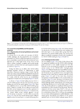
Figure 7. Confocal images of immunostained hCF-loaded bioprinted hydrogels on days 1, 7, and 14. Nucleus and keratocyte-specific protein (ALDH3A1)
are stained blue and green, respectively. Contrast: pure GelMA hydrogel. The scale bar is 100 μm.
3.5. In vivo biocompatibility and therapeutic in CECM-GelMA group (Figure 8B), which demonstrated
potential that the levels of corneal stromal haze and scarring were
3.5.1. Regeneration of corneal epithelium and corneal obviously higher in the corneal defect group than those
transparency observed in the CECM-GelMA group. The corneas were
To evaluate the in vivo safety and outcome of CECM- almost completely transparent 2 months after transplant of
GelMA hydrogel, the biocompatibility and therapeutic the CECM-GelMA hydrogels (Figures 8B and 9A).
potential of the CECM-GelMA hydrogel were studied in a
focal corneal defect model with the corneal defects of 5 mm 3.5.2. Histological observation
diameter and 1/3 the depth of the cornea. Focal corneal The HE staining (Figure 10A) results further showed that
defects without hydrogel transplant and normal groups the regenerated epithelium in the CECM-GelMA hydrogel
were used as controls. Schematic and surgical procedures group possessed a more regular arrangement than that
are shown in Figure 8A. observed in the control group. In addition, the superficial
corneal stroma in the CECM-GelMA hydrogel group
During the follow-up, all rabbits with CECM-GelMA
hydrogel transplant showed no inflammation or graft (Figure 10A) displayed regular structures similar to those in
normal corneas. However, both the cell layers of epithelium
dislocation. Fluorescein staining images revealed that the and the keratocytes in superficial stroma were significantly
defect area of the corneal epithelium gradually decreased over increased in the control group compared with the normal
time in the CECM-GelMA group and almost re-epithelialize group (Figure 10A part). At the same time, disordered
in 2 weeks, while the cornea was not yet re-epithelialize arrangement of superficial fibers in the corneal stroma was
completely in 4 weeks in the control group (Figure 9A and B).
observed (Figure 10A part). Moreover, the thickness of the
As shown in Figure 9B, the healing rate was 93.5% in corneal epithelial, stromal, and total thickness in the CECM-
CECM-GelMA group at 28 days post-operation, while the GelMA hydrogel group, control group, and normal group
healing rate was 84% in control group (Figure 9B). There was histologically assessed at 2 months after surgery (Figure
was a slight haze in the cornea of the CECM-GelMA group 10B). The results indicated no significant differences in the
from 2 weeks to 4 weeks, but all rabbits recovered with total thickness of the corneal among the groups. However,
favorable corneal transparency at 2 months, equivalent to compared with the normal tissue (24.87 ± 4.19 μm), the
the outcome of normal cornea (Figure 9A). Comparatively, control group exhibited a larger increase in the thickness of
the corneal defect group showed apparent corneal the corneal epithelial layer (52.71 ± 9.47 μm), demonstrating
scar formation during the 2 months follow-up periods the occurrence of heterogeneous re-epithelialization in the
(Figure 9A). The AS-OCT results showed higher reflected control group. Nevertheless, the thickness of the corneal
signal in corneal stromal layer in corneal defect group than epithelial layer of CECM-GelMA hydrogel group (30.40 ±
Volume 9 Issue 5 (2023) 485 https://doi.org/10.18063/ijb.774