
Page 495 - IJB-9-5
P. 495
International Journal of Bioprinting CECM-GelMA bioinks of DLP 3D printing for corneal engineering
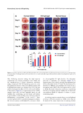
Figure 9. D-Luciferin experiment verifies epithelial regeneration after in vivo corneal defect. (A) Slit-lamp observations and fluoresce in staining images
of corneal defect, CG hydrogel-treated, and normal tissue at 0, 2, 4, and 8 weeks after surgery. (B) Quantitative results of corneal epithelial repair area in
in vivo model.
File). Following extensive testing, the ideal material is a biocompatible UV light absorber. The absorption
composition and printing parameters were found, and spectrum of tartrazine was measured (Figure S2 in
the manufacturing of corneal alternatives continued, Supplementary File), which indicated that tartrazine had
using a homemade DLP 3D bioprinting system to create obvious absorption in the UV band. The absorbance of the
the appropriate structure. Since most photoinitiators used absorbent at different concentrations was measured by a
in photopolymerization are cytotoxic, the CCK-8 kit was microplate reader (800TS, Bio Tek Instruments Inc., USA)
used to investigate the effect of 0.5% LAP on cell viability to obtain the molar absorption coefficient. Considering
in the system (Figure S4 in Supplementary File). GelMA both the printing layer and cell viability, the UV absorbent
samples cured with LAP have negligible effects on cell concentration should be adjusted to achieve a proper
viability after crosslinking, and the overall cell viability crosslinking depth.
is good within a short printing time (<60 min) and low
photoinitiator concentration (<0.5% (w/v)) . Then, According to previous reports, the accuracy of hydrogel
[39]
considering that GelMA is a relatively transparent solution, printing is difficult to determine. Unlike resins that can
the vertical accuracy is determined by tartrazine, which be inspected by contact inspection or scanning electron
Volume 9 Issue 5 (2023) 487 https://doi.org/10.18063/ijb.774