
Page 492 - IJB-9-5
P. 492
International Journal of Bioprinting CECM-GelMA bioinks of DLP 3D printing for corneal engineering
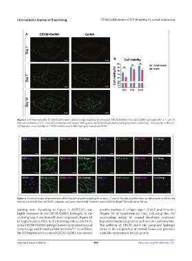
Figure 5. (A) Representative 3D live/dead stained confocal image of printed hCFs-loaded CECM-GelMA (CG) and GelMA hydrogels after 1, 7, and 14
days of incubation at 37°C. Live cells are stained with calcein-AM (green), and dead cells are stained with propidium iodide (red). The scale bar is 200 μm.
(B) Statistics of cell viability of CECM-GelMA and GelMA hydrogels loaded with hCFs.
Figure 6. Confocal images of immunostained hCF-loaded bioprinted hydrogels on days 1, 7, and 14. Nucleus, myofibroblast-specific protein (α-SMA) and
lumican are stained blue (by DAPI), magenta, and green, respectively. Contrast: pure GelMA hydrogel. The scale bar is 100 μm.
staining tests. According to Figure 7, ALDH3A1 was positive markers of collagen type I (Col-I) and vimentin
highly expressed in the CECM-GelMA hydrogels. In the (Figure S5 in Supplementary File), indicating that the
control group, it was basically down-expressed (Figure S8 surrounding matrix of corneal fibroblasts contained
in Supplementary File). In the following culture, the hCFs important functional proteins such as Col-I and vimentin.
in the CECM-GelMA hydrogel showed more physiological The addition of CECM made the composite hydrogel
morphology and formed cellular networks . In addition, closer to the composition of natural tissues and provided
[38]
the 3D-bioprinted structure of CECM-GelMA also showed a suitable environment for cell growth.
Volume 9 Issue 5 (2023) 484 https://doi.org/10.18063/ijb.774