Page 533 - IJB-9-5
P. 533
International Journal of Bioprinting                            GelMA/PEG-TA IPN networks for 3D bioprinting


            geometry (Discovery HR-1, TA Instruments) at 365 ±   90%, and 96% ethanol for 30 min each and 100% ethanol
            5 nm at room temperature. For all measurements, the gap   twice for 30 min. The constructs were dried with HMDS in
            between the plates was set to 500 µm.              a fume hood overnight. The samples were gold sputtered
                                                               using a Cressington Sputter coater 108 auto before imaging.
            2.5. Compressive and tensile tests
            Compression  stress-strain  analyses  were  performed  on   2.8. Degradation
            5 × 8  mm (height × diameter) cylindrical specimens in   Degradation of the photo-crosslinked and IPN hydrogels
            the wet state. Tests were performed at room temperature   was tested using 5 × 8 mm (height × diameter) cylindrical
            according  to ASTM 695 at  a compression  rate of 30%   specimens cast in a PDMS mold. Specimens were dried at
            per  min,  n = 5. The  compressive modulus (E c-mod ) was   37°C in an oven for 2 days to give the initial weight (m ).
                                                                                                            0
            determined at 10% compressive strain.              Then, specimens were separately submerged in 2 mL PBS
              Tensile  stress-strain  measurements  were performed   containing 2 U/mL collagenase at 37°C. Sodium azide
            according to ASTM D 638 using a ZwickRoell tensile   (0.02 wt%) was added to prevent bacterial growth. The
            tester. Samples with dumbbell shape (50 × 9 mm) in the   medium was exchanged every 2 days. After 6, 12, 24, 48,
            wet state were elongated at a speed of 10 mm/min at room   72, 96, 120, and 168 h, specimens (n = 3) were removed
            temperature. Starting from the initial position (30  mm   from the degradation media and washed with deionized
            grip-to-grip separation), the stress and elongation at break   water. Thereafter, the specimens were lyophilized to give
            of three samples of each gel were measured to obtain values   the dry weight (m ). The percentage of mass remaining
                                                                              d
            for the tensile modulus (E t-mod ) at 10% strain and elongation   (M ) was calculated according to Equation III:
                                                                 r
            at break ( max ). Tensile and compressive toughness were   m d
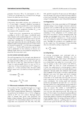
            used as parameters for the resistance to fracture of a   M =  m  ×100%                        (III)
                                                                    r
            hydrogel under stress and determined by integrating the      o
            area under the stress-strain curve.                2.9. 3D printing
            2.6. Gel content and water uptake                  All printing experiments were performed with an
            The swelling properties of the hydrogels were determined   extrusion-based 3D bioprinter (BioMaker 2.0, New
            by water uptake and gel content measurements based on   Jersey, SunP Biotech Inc., USA) using 6 wt% GelMA or
            gel weights in both swollen and dry states. The gels were   6 wt% GelMA/2 wt% 8PEGTA  solutions to which LAP
                                                                                        5
            first dried for 2  days (m ), then extracted in water for   as a photoinitiator was added at a concentration of 0.5
                                 0
            2 days (m ) and eventually dried in an oven at 37°C for   wt%. In addition, to the 6 wt% GelMA/2 wt% 8PEGTA
                                                                                                             5
                    s
            2 days (m ). All steps were performed at 37°C using three   solution, HRP was added at a final concentration of
                    d
            samples of 1 × 10 mm preformed disks. The water uptake   4 U/mL. The combination of a printing speed of 4 mm/s
            and gel content were calculated according to Equations I   and extrusion speed of 0.45 uL/s and needle diameter was
            and II:                                            optimized. Optimal results were  obtained  when needles
                                                               with a diameter of 25G (0.260 ± 0.019 mm, nominal inner
                           m                                   diameter) were used. Both larger and smaller diameter
               Gelcontent =  d  ×100                    (I)
                           m                                   needles did not give smooth and regular fibers. The
                            0
                                                               temperature of the nozzle and collecting plate was set at
                            m  − m                             room temperature in line with rheological temperature
               Water update =  s  d  ×100              (II)    sweep experiments. The printing speed and extrusion
                              m d                              speed were optimized by one-layer (10 × 10 × 0.25 mm
                                                               size, line distance of 2 mm) printing tests for both the 6
            2.7. Microscopic evaluation of the morphology      wt% GelMA and 6 wt% GelMA/2 wt% 8PEGTA  solutions
                                                                                                     5
            Cross-sections of the (degraded) hydrogels were imaged   (data not shown). The GelMA or GelMA/8PEGTA
                                                                                                             5
            by scanning electron microscopy (SEM) using a Jeol   solution at 37°C was transferred to a syringe (BD, 5 mL,
            JSM-IT100 Scanning Electron Microscope. Degradation   Luer-Lock tip) with dispensing needle. The syringe was
            was performed by incubation into a 2 U/mL collagenase   kept at 4°C for 10 min to form physical crosslinks followed
            solution at 37°C (see next section). Hydrogel samples   by 20 min at room temperature for equilibrium. At this
            taken  at  different  time  points  were  lyophilized  and   temperature, physical crosslinks remain present as shown
            broken in liquid nitrogen. The cell-printed hydrogels were   by the rheological data. On testing, with given GelMA or
            treated with 10% formaldehyde solution for 2 h at room   GelMA/8PEGTA solutions, a printing speed of 4  mm/s
                                                                             5
            temperature and dehydrated with 30%, 50%, 70%, 80%,   and extrusion speed of 0.45 uL/s were chosen for further

            Volume 9 Issue 5 (2023)                        525                         https://doi.org/10.18063/ijb.750

