
Page 536 - IJB-9-5
P. 536
International Journal of Bioprinting GelMA/PEG-TA IPN networks for 3D bioprinting
is the macromer concentration. Recent investigations and shows promising results as a matrix for cartilage
have shown that printing of GelMA can be performed at tissue regeneration, it shows a lower metabolic activity of
concentrations ranging from 5 wt% to 15 wt%, providing encapsulated human mesenchymal stem cells. In addition,
constructs with promising stability and cell viability [34,35] . it was also shown that co-crosslinking of the 8PEGTA
5
Moreover, it was shown that cell viability drastically with a hyaluronic acid tyramine conjugate improves
decreased with increasing GelMA concentration, while too the biological performance of the hydrogel. The lowest
low GelMA concentrations led to poor printing ability [36,37] . concentration of the 8PEGTA that can form a gel at low
5
In a previous study by Wang et al. , it was shown that enzyme and H O concentration was 2 wt%.
[43]
tyramine conjugated of 8PEGTA can be enzymatically 2 2
5
crosslinked using HRP as a catalyst in the presence of In preliminary experiments, it was shown that in
H O . The gel obtained at a concentration of 10 wt% is the temperature window just below the gel point of a 6
2
2
highly elastic. Although such a gel is cytocompatible wt% GelMA solution at 27°C, smooth and continuous
filaments could be extruded through a fine needle (25G).
At lower temperatures, the increasing gel strength led to
A
non-continuous, curled, and irregular filaments. At 30°C,
which is higher than the gel point, the inks were liquid-
like, and no continuous filaments were formed (Figure S1,
Supplementary File).
Similar experiments were performed on mixtures of
GelMA and the 8PEGTA . As reported in the previous
5
studies, depending on concentration, aqueous solutions of
gelatin or GelMA and PEG or PEG derivatives are turbid
indicating that phase separation has taken place [12,38,39] . This
B phase separation can lead to poor physical gelation of the
GelMA and consequently to non-printability of solutions
or bioinks. It was observed that solutions containing 6
wt% GelMA and 2 wt% 8PEGTA were transparent but
5
became translucent at higher 8PEGTA concentrations.
5
The light transmission of a 6 wt% GelMA/2 wt% 8PEGTA
5
solution in water at 680 nm was 85%, slightly lower
than that of a 6 wt% GelMA solution (89%). Moreover,
intensity plots from dynamic light scattering (DLS)
measurements at high dilution revealed the presence of
particles mainly with an average size of 70 nm (Figure S2,
Supplementary File). These data indicated the occurrence
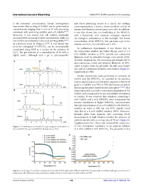
Figure 1. H-NMR spectra of GelMA (A, top) and gelatin (A, bottom) in of only microphase separation in an aqueous solution
1
D O and 8PEG (B, top) and 8PEGTA (B, bottom) in CDCl . of 6 wt% GelMA/2 wt% 8PEGTA . The gel point of this
2 5 3 5
A B
Figure 2. (A) Storage (G’) and loss modulus (G”) of 6 wt% GelMA and 6 wt% GelMA/2 wt% 8PEGTA physically crosslinked hydrogels as a function of
5
temperature. (B) Optimal printing temperature window based on tan δ value of 6 wt% GelMA and IPN.
Volume 9 Issue 5 (2023) 528 https://doi.org/10.18063/ijb.750